

Surgical Conditions & Procedures
An overview of assessment, treatment options and preparation for surgery
Robotic Surgery
Robotic surgery is a form of minimally invasive surgery in which the surgeon controls instruments from a console. A camera provides a three-dimensional view of the operating area; the system does not perform the operation independently.
Suitability depends on the diagnosis, anatomy, previous operations and the procedure required. Open and laparoscopic approaches are considered alongside robotic surgery.
Consultation covers the reasons for the proposed approach, alternatives, possible complications and the possibility of conversion to an open operation. Recovery depends on the operation and individual health; a robotic approach does not guarantee a shorter recovery or a better outcome.


Staging Laparoscopy
Staging laparoscopy examines the abdominal cavity through small incisions to look for disease that may not be visible on scans. It may be considered before selected operations for gastric, pancreatic or biliary cancer, or during assessment of peritoneal disease.
The surgeon inspects the abdominal surfaces and may take biopsies or peritoneal washings. These samples are examined for cancer cells and can provide additional information about the extent of disease.
Findings are considered alongside imaging and pathology at multidisciplinary review. They may change the proposed treatment, although a negative examination cannot exclude all microscopic disease. Anesthesia, procedural risks and the expected hospital stay are discussed beforehand.
Hepatobiliary & Pancreatic (HPB) Surgery
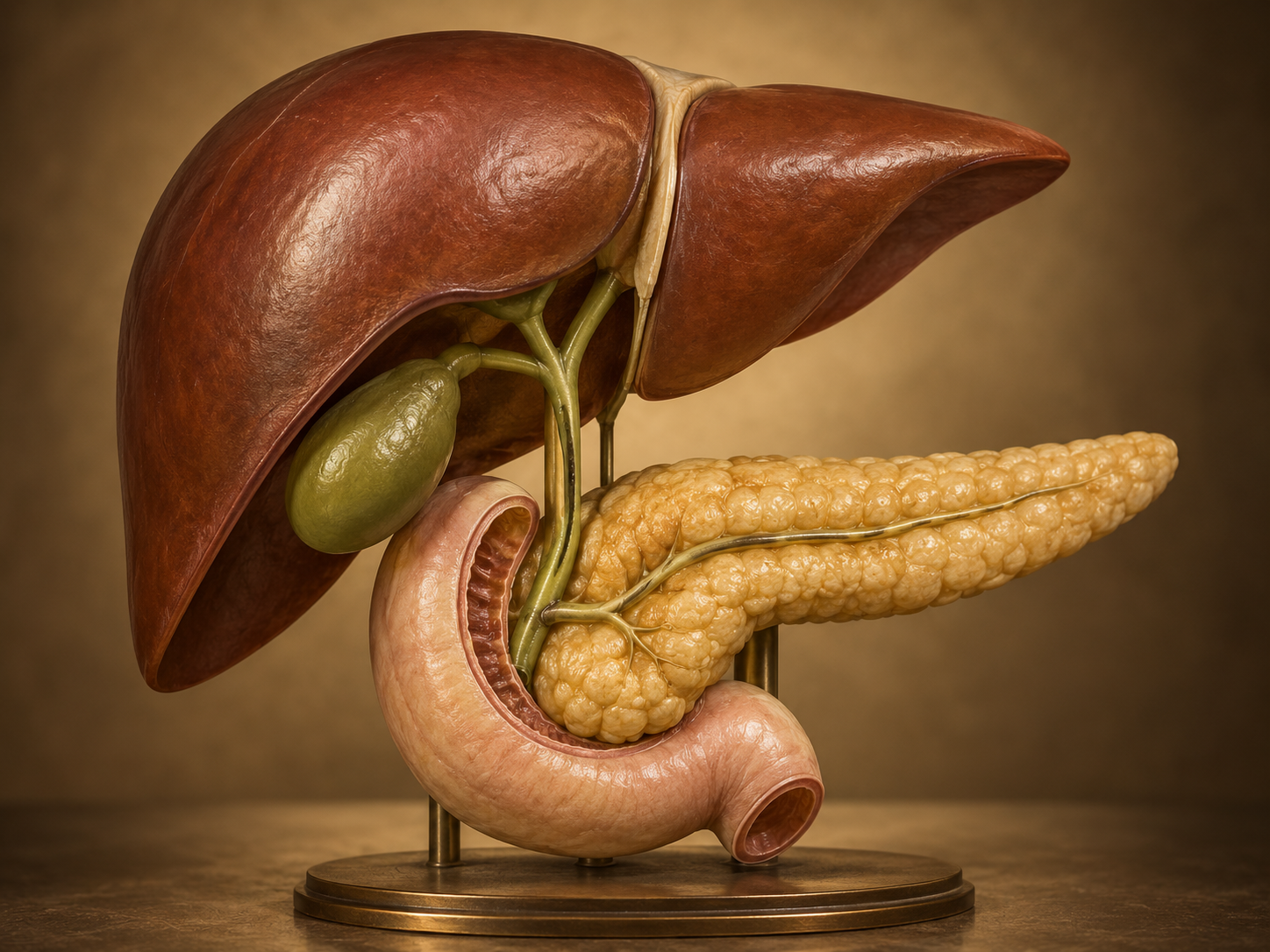
HPB surgery concerns the liver, pancreas, gallbladder and bile ducts. Conditions assessed include gallstones, bile-duct narrowing, liver and pancreatic cysts, pancreatitis, primary cancers and liver metastases.
Assessment may involve blood tests, CT, MRI/MRCP or endoscopic ultrasound. The diagnosis, extent of disease, liver function, nutrition and general health guide multidisciplinary discussion. Some conditions require monitoring or nonsurgical treatment.
When indicated, treatment may include gallbladder removal, liver resection or ablation, bile-duct reconstruction, or pancreatic surgery such as a Whipple procedure or distal pancreatectomy. The extent of treatment, possible complications and effects on digestion are explained before a decision is made.
Gastric Cancer & GIST Surgery
Gastric cancer and gastrointestinal stromal tumors (GIST) arise from different cell types and require different treatment plans. Assessment usually begins with endoscopy and tissue diagnosis, followed by imaging and additional staging tests when indicated.
Gastric cancer treatment may involve chemotherapy and removal of part or all of the stomach with associated lymph nodes. For GIST, surgery and the role of targeted medication depend on the tumor’s location, size and biological features.
Planning considers the extent of resection and the most appropriate operative approach. Nutritional support, changes in eating, possible complications and follow-up are discussed as part of preparation.


Colon Cancer & Benign Colorectal Disease
Colorectal assessment covers cancer, polyps unsuitable for endoscopic removal, diverticulitis, strictures, fistulas and selected inflammatory bowel conditions. Diverticulosis alone does not usually require an operation.
Investigations are chosen according to the problem and may include colonoscopy, biopsy and CT. Rectal cancer assessment may also require pelvic MRI and discussion of treatment before surgery.
An operation may remove a segment of bowel and associated lymph nodes when treating cancer. Open, laparoscopic and robotic approaches are considered individually. Consultation includes expected changes in bowel function, the risk of an anastomotic leak and whether a temporary or permanent stoma may be needed.
Sarcoma & Retroperitoneal Tumor Surgery
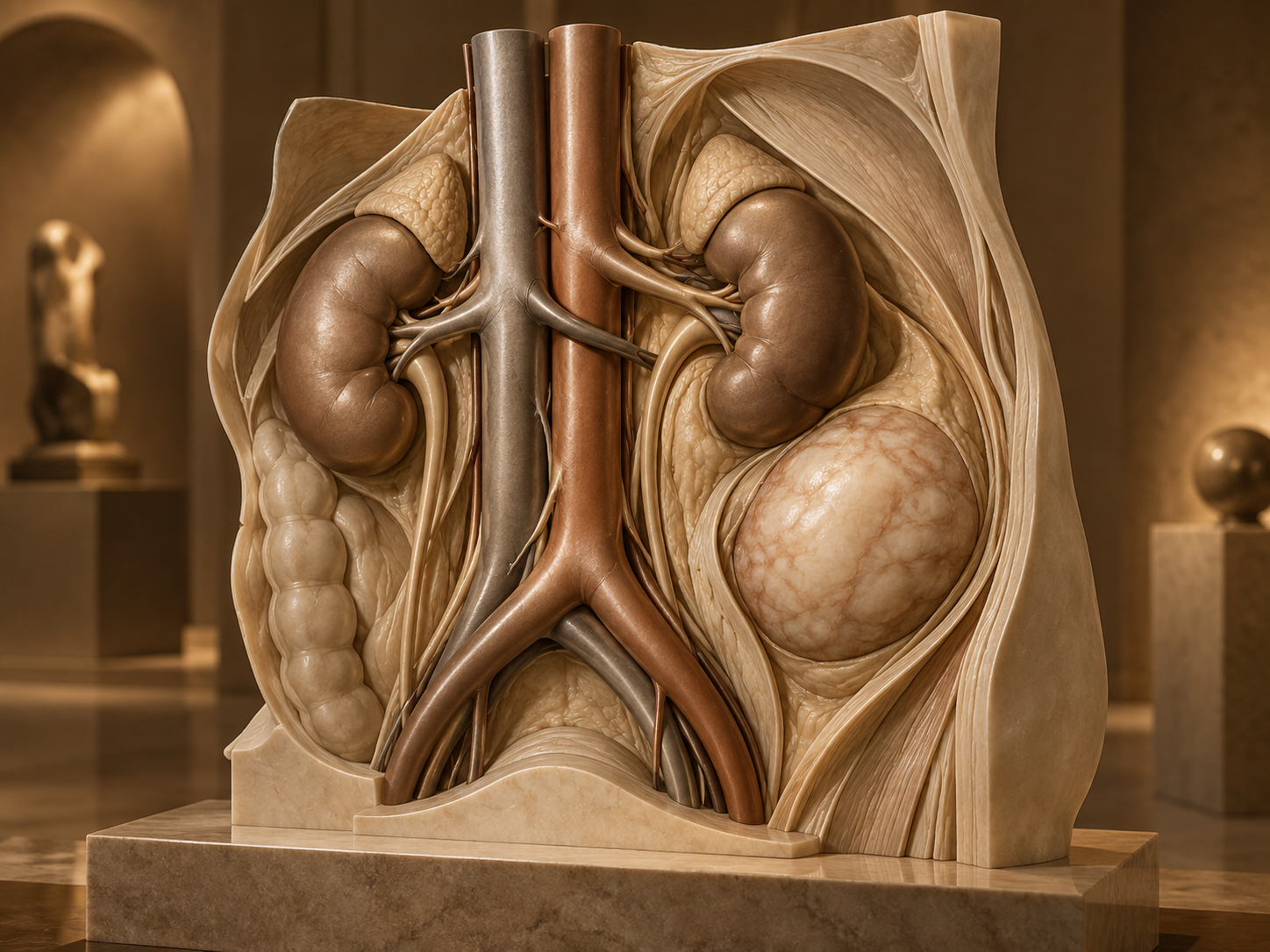
Sarcomas are uncommon tumors arising from connective tissues. In the abdomen and retroperitoneum—the space behind the abdominal cavity—they may lie close to organs, major blood vessels and nerves.
Assessment involves imaging, pathology review and, where appropriate, a planned image-guided biopsy. The tumor subtype and anatomical relationships guide discussion among surgery, radiology, pathology and oncology.
Surgery may require removal of adjacent organs to obtain an appropriate margin. The potential extent of resection, functional effects and complications are discussed in advance. Radiotherapy or systemic treatment may be considered in selected circumstances, and follow-up is planned according to the diagnosis.
Hernia & Abdominal Wall Surgery
A hernia occurs when tissue protrudes through a weakness in the abdominal wall. Assessment includes groin, umbilical, incisional, recurrent and parastomal hernias, with attention to symptoms, examination findings and previous repairs.
Treatment may involve observation or surgery, depending on the type of hernia and its risks. Repair can be open or minimally invasive and may use mesh; larger defects may require abdominal wall reconstruction.
Before surgery, discussion covers pain, recurrence, wound problems and mesh-related complications. Smoking, nutrition, weight and diabetes management may affect planning. Sudden severe pain, vomiting or a tender bulge that will not reduce requires urgent medical assessment.
Gallbladder Surgery (Cholecystectomy)
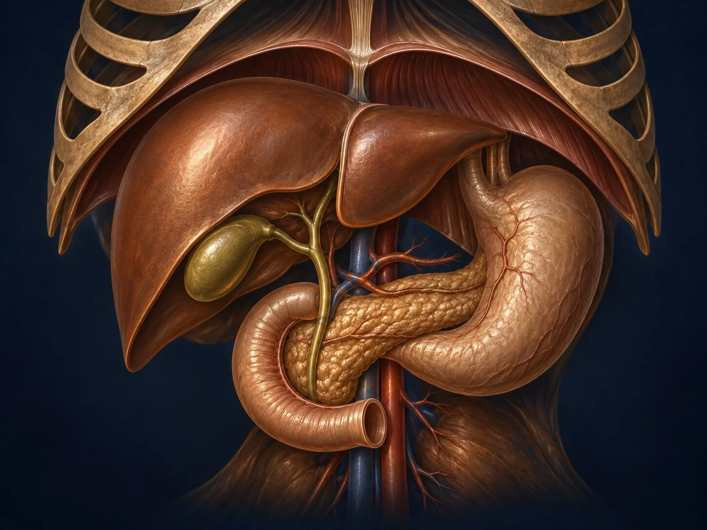
Gallbladder removal may be considered for symptomatic gallstones, inflammation and related complications. Stones found incidentally without symptoms do not always require surgery.
Assessment commonly includes ultrasound and blood tests. Further imaging or endoscopic treatment may be needed if stones in the bile duct are suspected. The timing of surgery depends on the diagnosis and clinical condition.
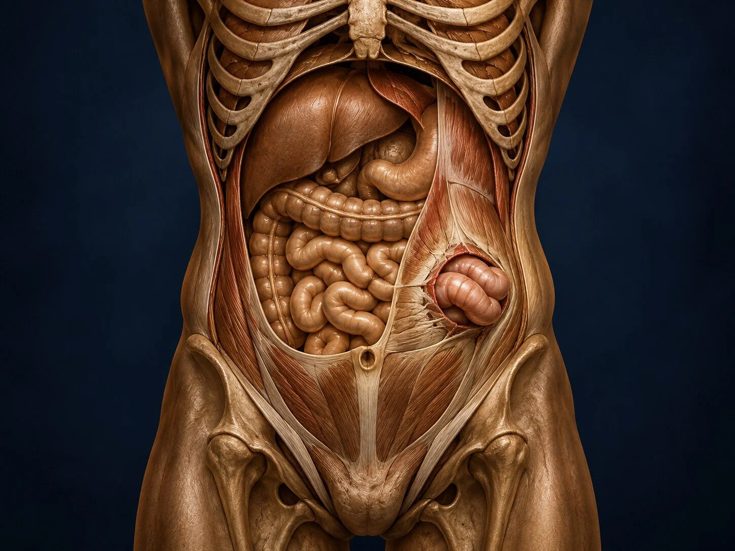
Cholecystectomy is often performed laparoscopically, although an open approach or a change of technique may be necessary. Discussion includes bleeding, infection, bile leakage and bile-duct injury. Hospital stay, return to activity and dietary advice are tailored to the operation and recovery.

Port-a-Cath (Central Venous Access) Insertion
An implanted venous port is a small reservoir beneath the skin connected to a central vein. It can provide access for repeated chemotherapy or other intravenous treatment when recommended by the treating team.
Placement is planned around the treatment schedule, medications and bleeding or infection risks. Local anesthesia, sedation and imaging guidance may be used according to the procedure and individual needs.
Possible complications include infection, thrombosis, bleeding and catheter problems. Patients receive instructions about wound care, access and maintenance, and when to seek review. Removal is considered when the port is no longer needed or if a complication requires it.
Gastrostomy Tube Placement
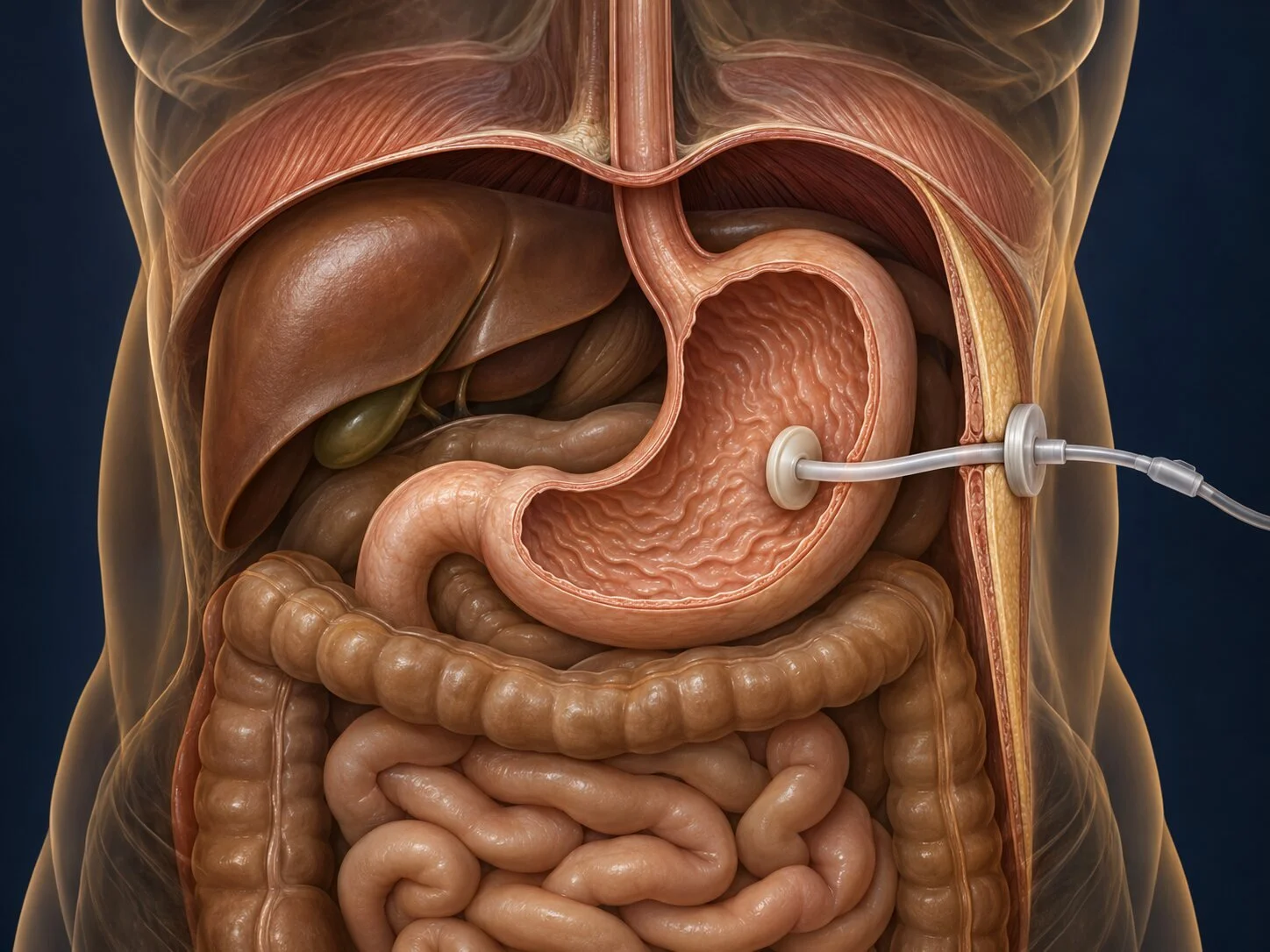
A gastrostomy tube provides access to the stomach when a person cannot meet nutritional needs by mouth. The decision considers the underlying condition, likely duration of feeding support and the person’s goals and preferences.
Placement may be endoscopic, radiological or surgical, depending on anatomy and clinical circumstances. Assessment involves the relevant medical team and dietitian, with swallowing specialists when appropriate.
Discussion covers alternatives and complications such as infection, leakage, displacement and aspiration. A gastrostomy does not eliminate aspiration risk. Feeding schedules, medication administration and tube care require an individual plan, with practical training for the patient or carers.
Adrenal Surgery
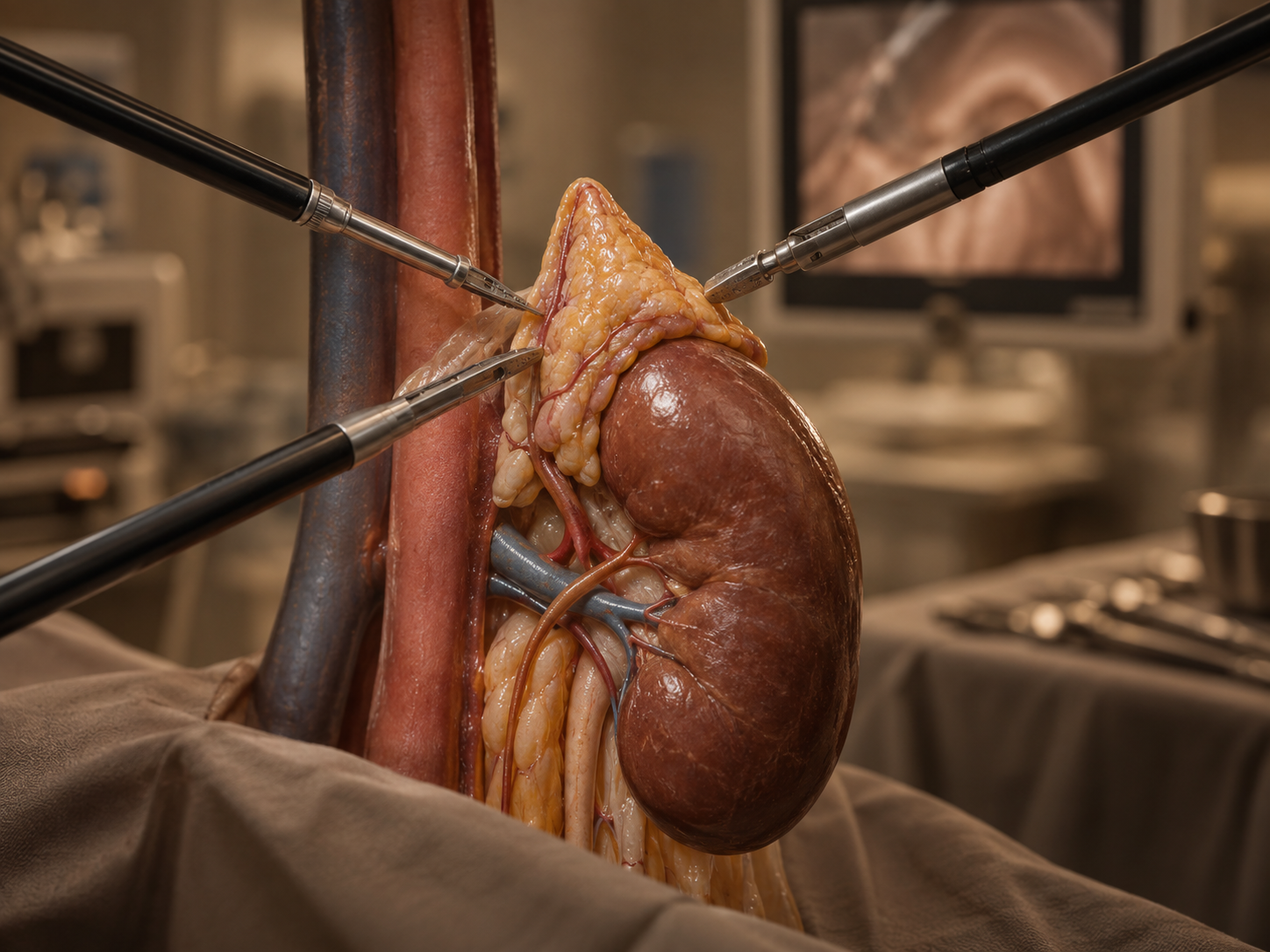
Adrenal masses may be found during scans for another condition. Assessment considers whether the lesion produces excess hormones and whether imaging suggests a benign or malignant process. Many adrenal findings do not require surgery.
Endocrinology review and appropriate hormonal tests help guide treatment. Some hormone-producing tumors require specific medical preparation before an operation.
When adrenalectomy is indicated, the approach depends on the size, imaging features and relationship to surrounding structures. Discussion includes surgical risks, potential changes in hormone production and whether temporary or ongoing hormone replacement may be necessary. Follow-up is coordinated with the relevant specialists.
Splenic Surgery
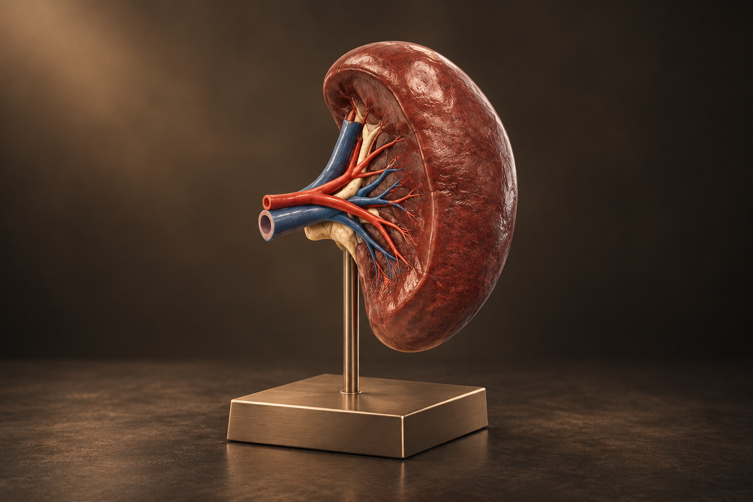
Splenic surgery may be considered for selected cysts, benign or malignant tumors, traumatic injury, symptomatic enlargement and certain blood disorders after specialist hematology review. Assessment focuses on the diagnosis, spleen size, symptoms, blood counts, imaging and whether preserving part or all of the spleen is feasible. When splenectomy is appropriate, minimally invasive surgery may be possible, while very large masses or complex disease can require an open approach. Because the spleen contributes to protection against infection, vaccination, antibiotic advice and prompt assessment of fever are important parts of planning and long-term care.Adrenal masses may be found during scans for another condition. Assessment considers whether the lesion produces excess hormones and whether imaging suggests a benign or malignant process. Many adrenal findings do not require surgery.
Endocrinology review and appropriate hormonal tests help guide treatment. Some hormone-producing tumors require specific medical preparation before an operation.
When adrenalectomy is indicated, the approach depends on the size, imaging features and relationship to surrounding structures. Discussion includes surgical risks, potential changes in hormone production and whether temporary or ongoing hormone replacement may be necessary. Follow-up is coordinated with the relevant specialists.
Lymph Node Dissection
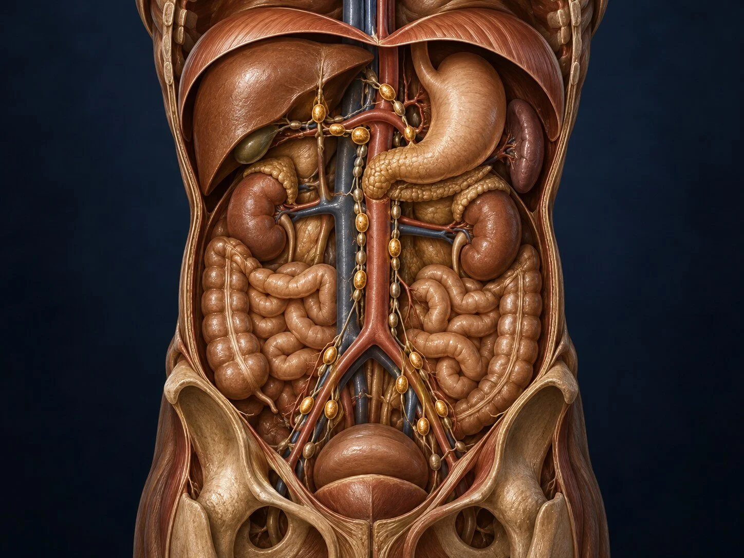
Lymph node dissection removes defined groups of nodes when cancer staging, local control or treatment requires it. The extent depends on the tumor type and location and may accompany surgery for gastric, colorectal, hepatobiliary, pancreatic, adrenal, sarcoma or other abdominal malignancies. Planning uses imaging, pathology and multidisciplinary discussion to balance oncologic benefit against risks such as bleeding, infection, lymphatic leakage, swelling and injury to nearby vessels or nerves. The operative findings and final pathology then help guide prognosis and any additional treatment.Lymph node dissection removes defined groups of nodes when cancer staging, local control or treatment requires it. The extent depends on the tumor type and location and may accompany surgery for gastric, colorectal, hepatobiliary, pancreatic, adrenal, sarcoma or other abdominal malignancies. Planning uses imaging, pathology and multidisciplinary discussion to balance oncologic benefit against risks such as bleeding, infection, lymphatic leakage, swelling and injury to nearby vessels or nerves. The operative findings and final pathology then help guide prognosis and any additional treatment.Adrenal masses may be found during scans for another condition. Assessment considers whether the lesion produces excess hormones and whether imaging suggests a benign or malignant process. Many adrenal findings do not require surgery.
Endocrinology review and appropriate hormonal tests help guide treatment. Some hormone-producing tumors require specific medical preparation before an operation.
When adrenalectomy is indicated, the approach depends on the size, imaging features and relationship to surrounding structures. Discussion includes surgical risks, potential changes in hormone production and whether temporary or ongoing hormone replacement may be necessary. Follow-up is coordinated with the relevant specialists.
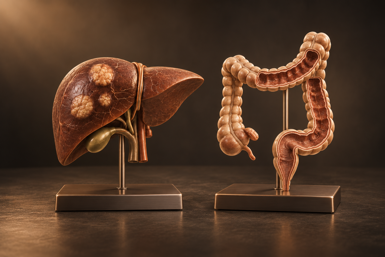
Liver Metastases & Combined HPB–Colorectal Surgery
Liver metastases most commonly arise from colorectal cancer but can also occur with other tumors. Assessment reviews the number and distribution of deposits, their relationship to major vessels and bile ducts, the condition of the remaining liver, disease outside the liver and response to systemic treatment. Treatment may include liver resection, ablation, chemotherapy or a staged combination, planned jointly with oncology, radiology and liver specialists. In selected patients, coordinated bowel and liver surgery can be performed together; in others, sequencing the operations improves safety and preserves future options.
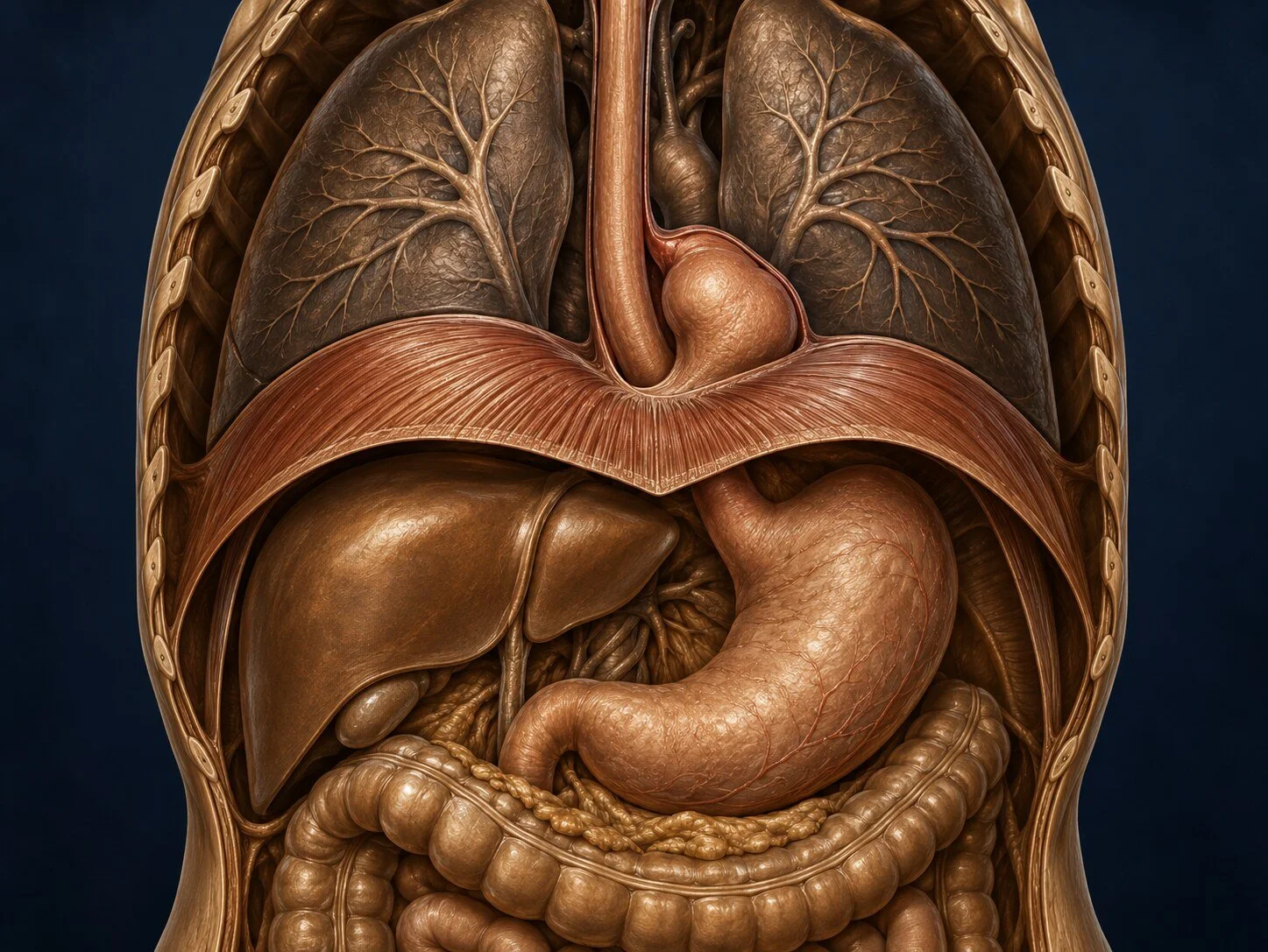
Diaphragmatic & Complex Hiatal Hernias
Diaphragmatic hernias allow abdominal organs to move through an abnormal opening in the diaphragm. They include large paraesophageal or recurrent hiatal hernias as well as traumatic and other acquired defects, and may cause reflux, swallowing difficulty, chest or upper-abdominal discomfort, breathlessness, anemia or obstruction. Assessment can require endoscopy, contrast studies, CT imaging and functional testing, tailored to the anatomy and symptoms. Repair returns organs to the abdomen, restores the diaphragmatic opening and may include an antireflux procedure or reinforcement; complex or recurrent cases require individualized planning because anatomy, scar tissue and previous mesh can change risk.
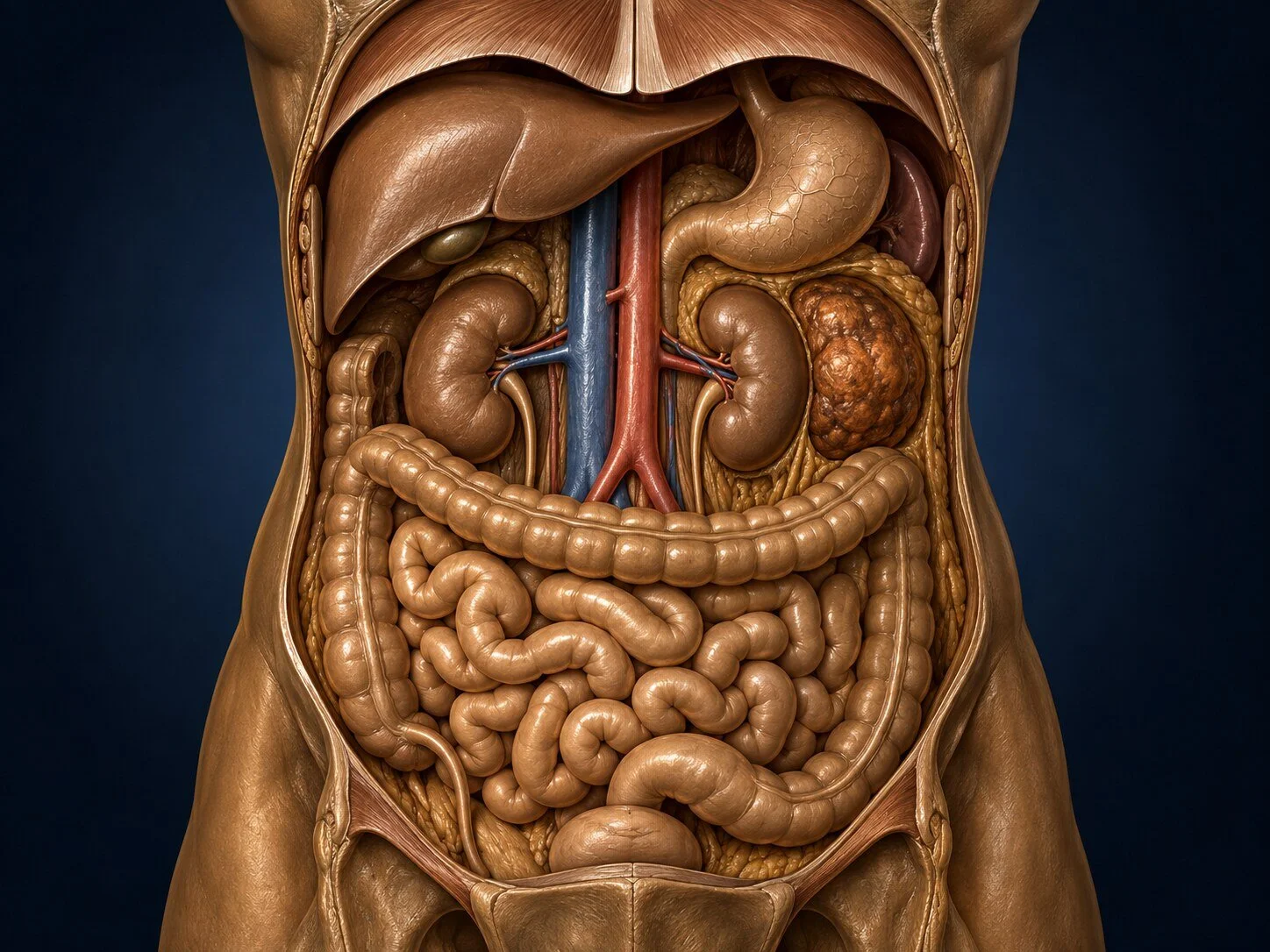
Abdominal & Retroperitoneal Masses
An abdominal or retroperitoneal mass may arise from an organ, soft tissue, lymph nodes, blood vessels or the lining of the abdominal cavity. The first priority is to define where it originates and whether it is inflammatory, benign or malignant without disrupting future treatment; biopsy is useful in selected cases but should be planned along an appropriate route. Assessment may combine contrast CT or MRI, specialist radiology and pathology review, blood tests and discussion in a multidisciplinary cancer meeting. Surgery can range from limited excision to multivisceral resection with vascular, urologic or other specialist support, and is recommended only when expected benefit justifies the functional and recovery risks.

Peritoneal Surface Malignancy & CRS–HIPEC
Peritoneal surface malignancy affects the lining of the abdominal cavity. Assessment considers the cancer type, distribution of disease, previous treatment, general health and whether a meaningful surgical benefit is realistic.
Cytoreductive surgery aims to remove visible disease and may involve several abdominal organs. HIPEC delivers heated chemotherapy within the abdomen during surgery. Its role and supporting evidence differ between cancer types; it is not appropriate for every patient with peritoneal disease.
A multidisciplinary discussion considers systemic treatment and other alternatives. Consultation addresses the extent of surgery, major complications, nutritional needs, possible stomas and the demands of recovery and follow-up.
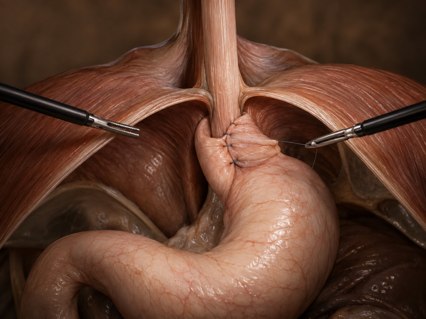
Hiatal Hernia & Antireflux Surgery
A hiatal hernia occurs when part of the stomach passes through the diaphragm. Assessment may be appropriate for persistent reflux, swallowing difficulties or a larger paraesophageal hernia, particularly when complications are suspected.
Endoscopy, contrast imaging and selected esophageal function tests help establish the cause of symptoms. Medication and lifestyle measures remain options for many people; surgery is considered after individual assessment.
Repair may include repositioning the stomach, closing the diaphragmatic opening and a fundoplication. Discussion covers alternatives, recurrent symptoms, difficulty swallowing, bloating and the possibility of further treatment. Eating and activity instructions are provided for recovery.
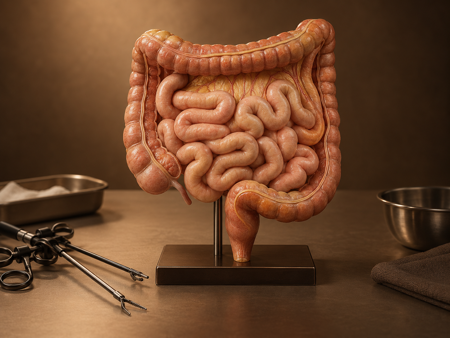
Small Bowel & Intestinal Surgery
Small-bowel surgery may be needed for tumors, obstruction, strictures, fistulas or compromised blood supply. Previous operations and inflammatory conditions can affect both diagnosis and treatment.
Assessment considers imaging, symptoms, nutritional status and the urgency of the problem. Some obstructions can be managed without surgery under clinical supervision; suspected bowel ischemia or perforation requires urgent assessment.
When surgery is necessary, the aim includes preserving as much functioning bowel as the condition allows. The proposed resection or reconstruction, risk of leakage, possible stoma and nutritional consequences are discussed. Complex fistulas may require nutritional support and staged treatment.
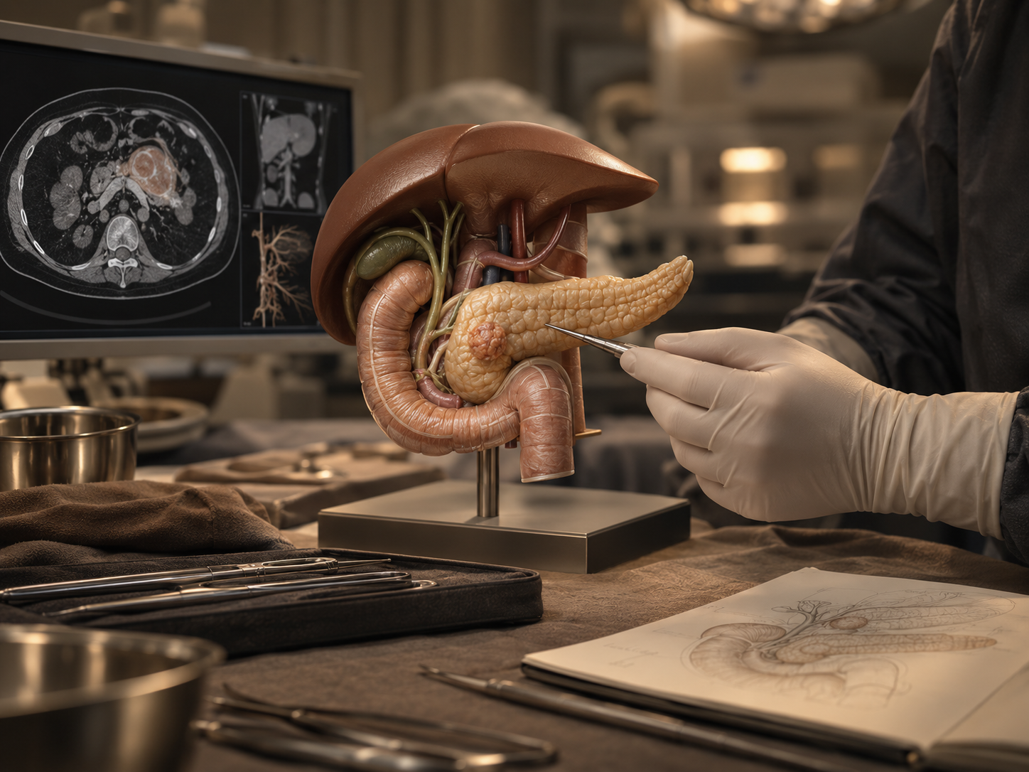
Neuroendocrine Tumor Surgery
Neuroendocrine tumors differ in their growth rate and ability to produce hormones. Treatment depends on the tumor’s origin, grade, stage and symptoms, rather than on its location alone.
Assessment may include pathology review, blood tests, CT or MRI and receptor imaging when indicated. Hormone-related symptoms may need treatment before an operation.
Options can include surveillance, surgery and medical or nuclear-medicine treatment. The extent of surgery is individualized, particularly for pancreatic or intestinal tumors and selected liver metastases. Multidisciplinary planning and follow-up involve the relevant surgical, oncology, endocrinology and nuclear-medicine teams.
Complex & Reoperative Abdominal Surgery
Repeat abdominal surgery may involve adhesions, altered anatomy, previous mesh, stomas or recurrent disease. Assessment begins with the current problem and a review of available imaging, pathology and previous operative records.
Planning considers nutrition, other medical conditions and the role of nonsurgical treatments. Endoscopy, interventional radiology and support from other specialties may be needed, and some conditions are best managed in stages.
Consultation sets out the intended benefit and limitations of further surgery, including organ injury, leakage, infection and possible stoma formation. The expected recovery and support after discharge are considered before deciding on treatment.
Consultation Information
Private and GeSY consultations are available at the German Medical Institute in Limassol. Nicosia appointments are also available at GMI’s outpatient offices within Zoi Medical Center (Makariou), 52 Archbishop Makarios III Avenue, 1075 Nicosia. Please specify your preferred location when booking.
☎ +357 25 208265
✉ charalambos.charalambous1@goc.com.cy
📍 German Medical Institute, 1 Nikis Avenue, Agios Athanasios, Limassol
◷ Monday – Friday, 08:00 – 16:00
Patient Information
For detailed preparation, diet, bowel-preparation and recovery guidance, please open Patient Information from the main navigation. Your individual written plan from the surgical and anesthesiology teams always takes priority.