

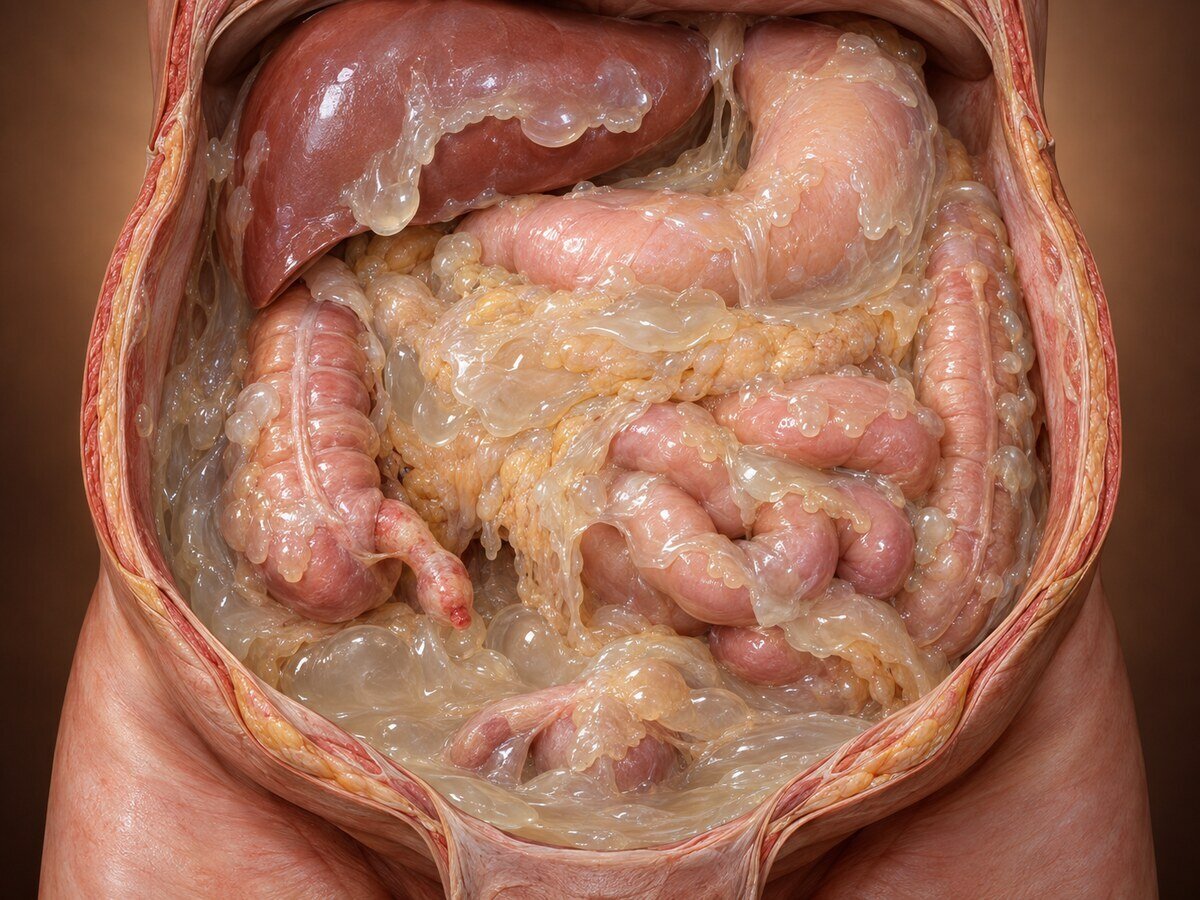
Pseudomyxoma peritonei is a rare condition in which mucus-producing tumour cells spread throughout the abdominal cavity and fill it progressively with jelly-like material. The historical name — "jelly belly" — describes the appearance accurately. It occupies an unusual position in oncology: the cells are often of low grade and rarely invade organs or spread through the bloodstream, yet the condition is fatal if untreated, because the accumulating mucus eventually compresses the bowel and the patient dies of obstruction and malnutrition. It is, in effect, a disease of relentless space-occupying growth rather than of destructive invasion.
That distinction is what makes it treatable. Because the disease stays within the abdominal cavity, an operation that removes everything within that cavity can be curative — and in specialist centres the great majority of patients with low-grade disease are alive many years later.
How it develops
The great majority arise from a mucinous neoplasm of the appendix. The sequence is mechanical and worth following, because it explains everything about the disease:
A mucinous tumour develops in the appendix and secretes mucus into its lumen.
The appendiceal lumen becomes obstructed and distends with mucus — a mucocele.
Pressure rises until the appendiceal wall perforates, often silently and without any acute illness.
Mucus containing tumour cells escapes into the peritoneal cavity.
The cells are carried by peritoneal fluid currents and distributed according to the redistribution phenomenon — a pattern unique to this disease. Because the cells do not adhere readily, they are swept along with the fluid and accumulate at the sites where peritoneal fluid is absorbed or where it pools: the greater omentum, the undersurface of the right diaphragm, the pelvis, the paracolic gutters, and the surfaces of the liver and spleen.
Crucially, the surfaces of the small bowel and its mesentery are relatively spared, because constant peristalsis keeps cells from settling there. This is the single anatomical fact that makes complete surgical removal possible.
The deposits continue to secrete mucus, which accumulates steadily, compressing bowel and eventually causing obstruction and nutritional failure.
Less commonly, pseudomyxoma arises from mucinous tumours of the ovary, colon, pancreas, urachus or gallbladder. In women, ovarian involvement is usually secondary to an appendiceal primary rather than the source, a point established by molecular studies and important because it changes the operation.
Classification
Grade determines outlook, and the terminology has been standardised:
Acellular mucin — mucus with no tumour cells; the best outlook.
Low-grade mucinous carcinoma peritonei (formerly disseminated peritoneal adenomucinosis) — abundant mucus with scanty, bland cells. The commonest, and the group with excellent long-term survival after complete treatment.
High-grade mucinous carcinoma peritonei — more cellular, with greater atypia and invasive features; a worse outlook.
High-grade with signet ring cells — the most aggressive, behaving like disseminated carcinoma.
Symptoms
The onset is insidious, and diagnosis is frequently delayed by years.
Progressive abdominal distension — the commonest presentation, often mistaken for weight gain, ascites from liver disease, or ovarian pathology
a new or enlarging hernia, with jelly-like material found in the sac at repair — a classical and diagnostic presentation
vague abdominal discomfort
appendicitis, or an appendix mass
an ovarian mass in women, frequently leading to gynaecological surgery before the true origin is recognised
change in bowel habit, early satiety
infertility
a substantial number are found incidentally at surgery or imaging for something else
Weight loss is typically absent until late, and patients often appear well despite a grossly distended abdomen — an incongruity that is itself characteristic.
How the diagnosis is made
CT of abdomen and pelvis — the key investigation. The appearances are distinctive: low-attenuation material throughout the abdomen; scalloping of the liver and splenic surfaces, where mucus indents the organ (a feature that distinguishes it from simple ascites, which does not indent); omental thickening; and, characteristically, central displacement of the bowel loops rather than the peripheral displacement seen with ordinary ascites. Calcification may be present.
MRI — useful for assessing small bowel and mesenteric involvement, which determines resectability.
PET-CT — of limited value, since low-grade mucinous disease is not metabolically avid. A negative PET is not reassurance here.
Tumour markers — CEA, CA 19-9 and CA 125 are useful for monitoring, with rising levels associated with recurrence.
Histology — essential for grading. A percutaneous biopsy is generally avoided because of the risk of seeding along the needle track.
An important practical point: when a mucocele or mucinous appendiceal tumour is found at appendicectomy, it should be removed intact without rupture, and the finding should prompt referral to a specialist peritoneal malignancy centre rather than simple discharge. Similarly, gelatinous material found unexpectedly in a hernia sac or at any operation should be sent for histology and the patient referred, not simply washed out.
Treatment
Cytoreductive surgery with HIPEC
This is the standard of care and it is potentially curative. The operation, developed and refined by Paul Sugarbaker, removes all disease-bearing peritoneal surfaces and organs while sparing the small bowel:
greater and lesser omentectomy, usually with splenectomy
stripping of the peritoneum from the right and left diaphragm
stripping of the pelvic peritoneum, with hysterectomy and bilateral oophorectomy in women
cholecystectomy and stripping of the lesser omentum and porta hepatis
right hemicolectomy or appendicectomy for the primary tumour
rectosigmoid resection where the pelvic disease requires it
stripping of the parietal peritoneum from the abdominal wall
preservation of the small bowel and its mesentery wherever possible — the limiting factor
Heated intraperitoneal chemotherapy, usually mitomycin C, is then circulated through the abdomen for sixty to ninety minutes to treat residual microscopic disease. Unlike in colorectal peritoneal metastases, the benefit of HIPEC in pseudomyxoma is well established.
The operation typically takes eight to twelve hours.
Results
For low-grade disease with complete cytoreduction, long-term survival in specialist centres is excellent — the majority of patients are alive at ten years, and many are cured. High-grade disease, and disease with signet ring cells, carries a considerably worse outlook, and this should be stated honestly.
The single strongest predictor of outcome is completeness of cytoreduction, which is why referral before any attempt at surgery elsewhere matters: previous incomplete operations create adhesions, distribute disease into the small bowel mesentery, and make subsequent complete removal harder.
Other approaches
Repeat cytoreductive surgery for recurrence, which is often worthwhile in low-grade disease.
Debulking surgery — removing mucus to relieve symptoms where complete removal is impossible; it does not cure but can give useful palliation for years in low-grade disease.
Systemic chemotherapy — of limited benefit in low-grade disease, which divides slowly; more useful in high-grade disease.
PIPAC — aerosolised intraperitoneal chemotherapy, used in selected patients with unresectable disease.
Recovery
Hospital stay is typically two to three weeks, with a period in critical care. Complications occur in a significant proportion. Recovery to normal activity takes three to six months. After splenectomy, vaccination and antibiotic advice are lifelong. Women undergoing hysterectomy and oophorectomy require discussion of hormone replacement and, where relevant, fertility preservation beforehand.
Follow-up
Lifelong, with clinical review, tumour markers and CT at intervals, since recurrence may occur many years later and is often treatable.
When to seek an opinion
Referral to a specialist peritoneal malignancy centre is indicated for: any mucinous appendiceal tumour or appendiceal mucocele; gelatinous material found in a hernia sac or at any abdominal operation; unexplained mucinous ascites; and a CT showing scalloping of the liver surface with central displacement of bowel. Referral should precede any attempt at surgical treatment, because the first operation is the one with the best chance of being complete.
Common questions
Is this cancer?
It is a form of cancer, but it behaves unlike most. The cells are often low grade and do not usually invade organs or spread through the bloodstream to the lungs or liver. The problem is that they keep producing mucus, which accumulates and eventually presses on the bowel. So it is dangerous through sheer accumulation rather than through invasion, and that is precisely why an operation that removes it all can cure it.
Where does it come from?
Nearly always from a small tumour in the appendix, which becomes blocked, fills with mucus and eventually splits — often without causing any noticeable illness. The cells then spread around the abdominal cavity in the natural fluid currents. In women it is often first noticed as an ovarian mass, but the ovary is usually affected secondarily rather than being the source.
Why does the disease spare the small bowel?
Because the small bowel is constantly moving, which prevents the cells from settling on it, while they accumulate at the sites where peritoneal fluid pools or is absorbed. This is a fortunate feature of the disease, since the small bowel cannot be removed — it is what makes complete surgical clearance possible.
How big is the operation?
Large — typically eight to twelve hours, removing the lining of the abdomen and several organs that can be spared, followed by heated chemotherapy circulated through the abdomen. Hospital stay is two to three weeks, often with time in intensive care, and full recovery takes three to six months. It is done in a small number of specialist centres.
What are my chances?
For low-grade disease that is completely removed, they are good — the majority of patients are alive ten years later and many are cured. For high-grade disease, and particularly where signet ring cells are present, the outlook is considerably less favourable. The grade from the microscope examination is the key piece of information, and you should ask for it directly.
Why should I be referred before any operation?
Because the first operation has the best chance of removing everything, and that completeness is the strongest predictor of long-term survival. Previous incomplete surgery creates scarring and can spread disease onto the small bowel, making later complete removal harder or impossible. If mucus or a mucinous appendix tumour has been found, referral should come before further surgery, not after.
Can it come back?
Yes, sometimes many years later, which is why follow-up is lifelong. Recurrent low-grade disease can frequently be treated again with a further operation, and repeat surgery is worthwhile in selected patients.
Does chemotherapy help?
Heated chemotherapy given into the abdomen at the time of surgery is an established part of the treatment for this condition. Conventional chemotherapy given into a vein is of limited value for low-grade disease, because the cells divide slowly and chemotherapy works mainly on rapidly dividing cells. It has more of a role in high-grade disease.
Related conditions
Other conditions of the peritoneal disease covered on this site:
This page provides general information and does not replace an individual medical consultation. Pseudomyxoma peritonei should be assessed in a specialist peritoneal malignancy centre.
