

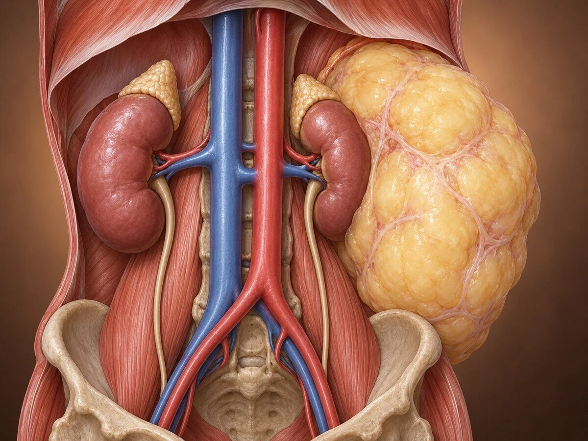
Retroperitoneal liposarcoma is a malignant tumour of fat cells arising in the space behind the abdominal cavity. It is the commonest retroperitoneal sarcoma. It has two characteristics that set it apart from most abdominal cancers: it frequently grows to an enormous size before causing any symptom at all — tumours weighing several kilograms are not unusual at diagnosis — and it rarely spreads to distant organs. The threat is local. It recurs where it started, repeatedly, and the aim of treatment is therefore local control, achieved above all by a properly planned first operation.
That last point deserves emphasis at the outset: the first operation is the one that matters most. The difference in long-term outcome between a well-planned resection in a specialist sarcoma centre and an unplanned excision elsewhere is one of the largest and most consistent findings in this field.
How it forms
Liposarcoma arises from primitive mesenchymal cells that differentiate along adipocytic lines. The retroperitoneum is its commonest site because it contains a large volume of loose fat and, critically, because it can accommodate a great deal of growth before anything is compressed.
The subtypes are genetically distinct, and the genetics determine the behaviour:
Well-differentiated liposarcoma (atypical lipomatous tumour) — the commonest. It is characterised by amplification of the 12q13-15 region, producing supernumerary ring or giant marker chromosomes containing multiple copies of MDM2 and CDK4. MDM2 is the natural inhibitor of p53; amplifying it effectively switches off the cell's principal tumour suppressor without mutating it. The tumour looks like mature fat with scattered atypical cells. It does not metastasise, but it recurs locally and relentlessly.
Dedifferentiated liposarcoma — arises when a well-differentiated tumour acquires additional genetic changes and a component transforms into a high-grade, non-lipogenic sarcoma. This occurs in a substantial proportion over time, and it is why a long-standing "benign-looking" retroperitoneal fatty mass cannot simply be ignored. The dedifferentiated component can metastasise, usually to the lung. On imaging, the characteristic appearance is a large fatty mass containing a discrete solid nodule — and that nodule is the dangerous part.
Myxoid liposarcoma — driven by the FUS-DDIT3 fusion gene. Far commoner in the limbs than the retroperitoneum. It has a distinctive pattern of spread to other soft tissue sites, bone and the spine rather than the lung, which changes how it is staged and followed.
Pleomorphic liposarcoma — rare, high grade, genetically complex, and aggressive.
The MDM2 amplification test (by FISH or immunohistochemistry) is diagnostically decisive: it distinguishes a well-differentiated liposarcoma from a benign lipoma, which look very similar under the microscope but have entirely different implications. A large retroperitoneal "lipoma" should be regarded as a liposarcoma until MDM2 testing says otherwise.
Symptoms
The retroperitoneum is a forgiving space, and tumours are frequently very large before anything declares itself.
A painless, gradually enlarging abdominal mass — often noticed by the patient as increasing abdominal girth or clothes not fitting, and sometimes attributed to weight gain
vague abdominal or back discomfort, and a sense of fullness
early satiety
leg swelling, from venous or lymphatic compression
neurological symptoms — pain, numbness or weakness in a leg from nerve compression or invasion
urinary symptoms, or hydronephrosis found incidentally, from ureteric compression
a hernia, or scrotal swelling
weight loss is characteristically absent until late, which is one reason the diagnosis is delayed
a significant proportion are found incidentally on imaging performed for something else
How the diagnosis is made
CT of chest, abdomen and pelvis with contrast — the primary investigation. It defines the size and extent, the relationship to kidney, ureter, major vessels, pancreas, bowel and nerve roots, and assesses for metastases. Fat density within the mass is highly suggestive of liposarcoma.
MRI — superior for assessing the relationship to nerve roots, muscle and the spinal canal, and for characterising tissue.
Image-guided core biopsy — this should be discussed carefully. Historically biopsy was avoided for fear of seeding. Current practice in specialist centres is that a coaxial core biopsy, taken through a retroperitoneal approach that will be included in the resection specimen, is appropriate and often important, because it establishes the subtype and grade, distinguishes sarcoma from lymphoma, germ cell tumour and metastatic disease — which are treated entirely differently and not by surgery — and allows consideration of treatment before surgery. Transperitoneal biopsy routes are avoided.
MDM2 testing — essential to distinguish liposarcoma from lipoma.
Assessment of renal function and, where nephrectomy may be required, split renal function, since resection of one kidney is frequently part of the operation.
The key principle: a retroperitoneal mass should be referred to a specialist sarcoma centre before biopsy or surgery, not after. Unplanned excision by a surgeon unfamiliar with these tumours compromises the one opportunity for a well-executed first resection.
Treatment
Surgery
Complete surgical resection is the only curative treatment, and its quality determines everything.
Modern practice is compartmental or extended resection: rather than simply shelling the tumour out of its pseudocapsule — which leaves microscopic disease behind, since tumour cells extend beyond the visible margin — the tumour is removed together with the surrounding fat and any adjacent organs that abut it, to obtain a true margin of normal tissue. In practice this commonly means removing en bloc the ipsilateral kidney and adrenal, the colon on that side, sometimes the tail of the pancreas and spleen, and portions of the psoas or abdominal wall muscle.
This is more extensive than an enucleation, and it is justified by better local control — which in a disease that kills through local recurrence translates into better survival. It is also the reason these operations belong in high-volume centres with vascular, urological and hepatobiliary support available.
Where the tumour involves major vessels, vascular resection and reconstruction is undertaken in specialist units. Involvement of the spine, extensive bilateral disease, or encasement of the superior mesenteric artery may render a tumour unresectable.
Radiotherapy
Preoperative radiotherapy has been the subject of the major trial in this field, STRASS, which found no overall improvement in abdominal recurrence-free survival when added to surgery. Subsequent analysis suggested a possible benefit in well-differentiated and low-grade dedifferentiated liposarcoma, and no benefit — possibly harm — in leiomyosarcoma. Practice therefore varies, and radiotherapy is used selectively rather than routinely. Patients should be told that this is an area of genuine uncertainty rather than settled practice.
Systemic therapy
Chemotherapy has a limited role. Doxorubicin-based regimens, with ifosfamide or dacarbazine, are used for high-grade or metastatic disease. Trabectedin and eribulin are active in liposarcoma specifically. MDM2 inhibitors and CDK4 inhibitors, targeting the defining genetic abnormality directly, are in clinical trials and represent the most promising direction.
Recurrence
Local recurrence is common, particularly in well-differentiated disease, and repeat resection is frequently worthwhile — patients may undergo several operations over many years with good quality of life. The decision to operate again is weighed against the pattern of growth: rapidly growing, multifocal recurrence is generally not helped by further surgery, while a solitary slow-growing recurrence often is.
Recovery
These are major operations. Hospital stay is typically one to two weeks, often with a period in critical care, and recovery to full activity takes two to three months. Where a kidney has been removed, renal function is monitored and care taken with nephrotoxic drugs. Where a section of bowel has been resected, function may change. Leg weakness or numbness may follow sacrifice of a nerve, and should be discussed in advance where it is anticipated.
Follow-up
Long-term and protocol-driven, since recurrence may occur many years after resection. CT of the abdomen and pelvis at intervals, with chest imaging for the subtypes that metastasise. Follow-up continues for at least ten years, and in well-differentiated disease effectively indefinitely.
When to seek an opinion
Any retroperitoneal or abdominal mass without an obvious explanation, particularly a fatty mass, should be referred to a specialist sarcoma centre before biopsy or attempted removal. A "lipoma" in the retroperitoneum is not a lipoma until proved so by MDM2 testing.
Common questions
How did it get so large without me noticing?
Because the space behind the abdominal cavity is loose and accommodating, so a tumour can expand for years, displacing organs gradually rather than compressing them suddenly. Many people notice only increasing abdominal size, which is easily put down to weight gain. It is a feature of the disease, not something you missed.
Is this the same as a lipoma?
Under the microscope they can look very similar, which is exactly why a specific genetic test is done. A true lipoma is harmless; a well-differentiated liposarcoma looks almost the same but recurs and can eventually transform into a more aggressive tumour. A large fatty mass in this part of the body is treated as a sarcoma until that test says otherwise.
Has it spread?
Usually not, and this is an important difference from most abdominal cancers. The commonest types rarely spread to other organs. The problem is that they come back in the same place. That is why so much emphasis is placed on removing them completely the first time.
Why does the operation involve removing my kidney and part of my bowel?
Because simply peeling the tumour out leaves microscopic disease behind in the surrounding tissue, and that is what regrows. Removing the tumour together with the fat and the adjacent organs it abuts gives a genuine margin of normal tissue and considerably better long-term control. One kidney is enough for normal life, and kidney function is checked beforehand.
Why should I be treated at a specialist centre?
Because the first operation is the single biggest determinant of long-term outcome, and the difference between a properly planned resection and an unplanned one is substantial. These tumours are uncommon, the operations are complex and often need vascular and urological expertise on hand, and the results in high-volume centres are consistently better. Asking for that referral is entirely reasonable.
Should I have a biopsy first?
Often yes, taken through a carefully chosen route by a specialist centre. It establishes the exact type and grade, and it rules out other conditions that look similar on scans — such as lymphoma or a germ cell tumour — which are treated with drugs rather than surgery. Older advice to avoid biopsy has been largely revised, though the route matters.
Do I need radiotherapy?
This is an area where the evidence is genuinely unsettled. A large trial did not show an overall benefit from radiotherapy before surgery, though there may be benefit for particular subtypes. It is therefore used selectively, and you should expect a discussion about the uncertainty rather than a firm recommendation either way.
Will it come back?
It may, and possibly more than once over many years. That is the nature of this disease. The good news is that recurrence is often slow and can frequently be removed again, and many patients live for a long time with good quality of life through repeated operations. This is why follow-up scans continue for a decade or more.
How long is the recovery after surgery for retroperitoneal liposarcoma?
These are major operations. The hospital stay is typically one to two weeks, often including time in intensive care, and full recovery takes two to three months.
Related conditions
Other conditions of the sarcoma & retroperitoneal covered on this site:
This page provides general information and does not replace an individual medical consultation. Retroperitoneal sarcoma should be assessed in a specialist sarcoma centre before biopsy or surgery.
