

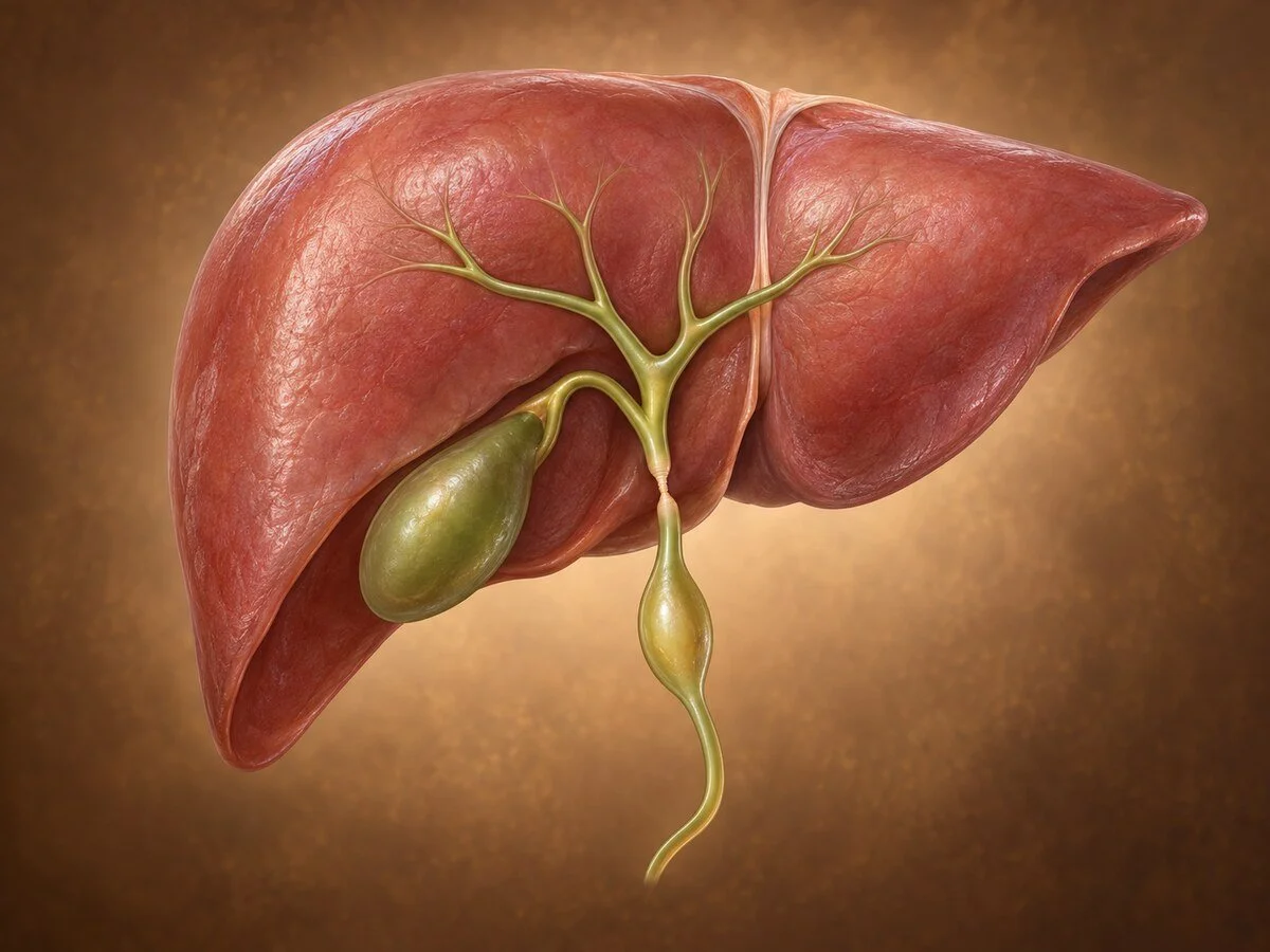
Not every narrowing or swelling of the bile duct is cancer. Benign strictures, congenital cysts and rare benign tumours all produce obstruction and jaundice, and all can resemble malignancy closely on imaging. Establishing which is present matters, because the treatments differ entirely — and because some benign conditions carry a long-term cancer risk of their own that dictates how they are managed.
Benign bile duct strictures
How they form
A stricture is a fibrous narrowing. Scar tissue forms wherever the duct has been injured, and the bile duct is particularly vulnerable because its blood supply runs in a narrow axial pattern along the wall, chiefly at the three and nine o'clock positions. Interrupt that supply and the duct heals badly and scars readily. The causes reflect this:
Surgical injury, most often during cholecystectomy — the commonest cause, dealt with on its own page.
Chronic pancreatitis — fibrosis in the pancreatic head compresses the intrapancreatic bile duct.
Primary sclerosing cholangitis — immune-mediated inflammation producing multiple strictures and dilatations, giving the characteristic beaded appearance.
IgG4-related sclerosing cholangitis — part of a systemic autoimmune condition, often accompanying autoimmune pancreatitis. It matters disproportionately because it responds to steroids, and it mimics cholangiocarcinoma closely enough that patients have undergone major resection for what was treatable inflammation.
Recurrent stones and chronic biliary infection.
Ischaemia — after liver transplantation or hepatic arterial injury.
Mirizzi syndrome — a stone impacted in the gallbladder neck compressing, and occasionally eroding into, the common hepatic duct.
Assessment
The dominant question is always whether the stricture is malignant. MRI with MRCP defines the anatomy and the length and position of the narrowing. Endoscopic ultrasound with biopsy, and brushings or cholangioscopy with directed biopsy at ERCP, provide tissue. Serum IgG4 is measured wherever autoimmune disease is plausible.
The honest position is that a proportion of strictures cannot be confidently classified before operation. Some patients undergo resection for disease that proves benign. That is an accepted consequence of not missing a curable cancer, and patients are entitled to have it explained in exactly those terms rather than discovering it afterwards.
Treatment
Endoscopic treatment is first-line for most benign strictures — dilatation with placement of multiple side-by-side plastic stents, or a fully covered metal stent, exchanged over several months. This succeeds in the majority. Surgical reconstruction, usually Roux-en-Y hepaticojejunostomy joining healthy duct above the stricture to a limb of jejunum, is used where endoscopic treatment fails, where the stricture is complete, or after significant injury. IgG4-related disease is treated medically with steroids.
Choledochal cysts
How they form
Congenital dilatations of the biliary tree. The leading explanation is an anomalous pancreaticobiliary junction, in which the pancreatic and bile ducts unite outside the duodenal wall, beyond the control of the sphincter of Oddi. Pancreatic enzymes reflux freely into the bile duct, and chronic exposure to activated enzymes damages the wall, weakening it so that it dilates.
That same chronic chemical injury drives metaplasia and dysplasia in the duct lining, which is why choledochal cysts carry a substantial lifetime risk of cholangiocarcinoma — a risk that rises with age and that persists if the cyst is merely drained rather than removed. This single fact governs their management.
The Todani classification describes five types by the pattern of dilatation and determines the operation. Type I, fusiform dilatation of the extrahepatic duct, is much the commonest. Type V, multiple intrahepatic cysts, is Caroli disease.
Symptoms
Children classically present with jaundice, abdominal pain and a palpable mass, though all three together are uncommon. Adults present with pain, recurrent jaundice, cholangitis or pancreatitis, or are found incidentally. Many are now identified on antenatal ultrasound.
Treatment
Complete excision of the cyst with hepaticojejunostomy reconstruction. The principle that matters: internal drainage procedures, performed historically, are no longer acceptable, because leaving abnormal epithelium in place leaves the cancer risk in place. Surveillance continues after excision, since risk persists in any retained ductal tissue. Caroli disease confined to one part of the liver may require hepatectomy; diffuse disease may ultimately require transplantation.
Benign tumours of the bile duct
Rare. Biliary adenoma, granular cell tumour and biliary papillomatosis all occur. Biliary papillomatosis is the one to recognise: multiple papillary lesions throughout the ducts producing mucin that obstructs bile flow, with a high rate of malignant transformation. It is treated as a premalignant condition requiring resection where feasible.
Because benign biliary tumours cannot reliably be distinguished from cholangiocarcinoma before operation, they are generally resected rather than observed.
Recovery
After hepaticojejunostomy, hospital stay is typically seven to ten days with recovery over six to eight weeks. Bile leak and later anastomotic stricture are the specific complications. Patients with a biliary-enteric anastomosis remain susceptible to cholangitis lifelong and should seek attention promptly with fever and rigors.
Follow-up
Long-term for all of these. Liver function tests and imaging detect recurrent stricture; for choledochal cysts and papillomatosis, surveillance addresses the continuing malignancy risk.
Common questions
My scan shows a narrowing in the bile duct. Does that mean cancer?
Not necessarily. Narrowings can result from previous surgery, chronic pancreatitis, inflammation or infection as well as from a tumour. Because the distinction is genuinely difficult and cancer must not be missed, these are investigated thoroughly, and occasionally the answer is only certain once the affected segment has been removed.
What is a choledochal cyst and why does it need removing rather than draining?
It is a congenital swelling of the bile duct, usually caused by an abnormal junction between the bile and pancreatic ducts that lets digestive enzymes wash back into the bile duct. The lining is chronically damaged and carries a significant lifetime risk of developing cancer. Draining the cyst relieves symptoms but leaves that lining, and therefore the risk, in place.
I had a cyst drained years ago rather than removed. Does that matter?
Yes, and it is worth being reviewed. Older operations drained these cysts internally. Current practice is complete excision, and patients treated the older way are generally advised to have specialist assessment because the cancer risk continues.
Can a bile duct stricture be treated without an operation?
Often, yes. Most benign strictures are managed endoscopically, by stretching the narrowing and placing stents that are exchanged over several months. Surgery is used when that fails, or when the duct is completely blocked.
What is IgG4 cholangitis?
An autoimmune condition that inflames and narrows the bile ducts and closely resembles bile duct cancer on scans. It matters because it responds to steroid treatment rather than surgery. A blood test for IgG4 forms part of the assessment when the cause of a stricture is unclear.
Why do I keep getting infections after my bile duct reconstruction?
When the bile duct is joined directly to the bowel, the valve that normally separates them is gone, so bacteria travel upward more easily. Episodes of fever with shivering should be treated promptly. Recurrent episodes may mean the join has narrowed and should be investigated.
Related conditions
Other conditions of the gallbladder & bile ducts covered on this site:
This page provides general information and does not replace an individual medical consultation. Assessment and treatment are decided for each patient after review of their history, examination and imaging.
