

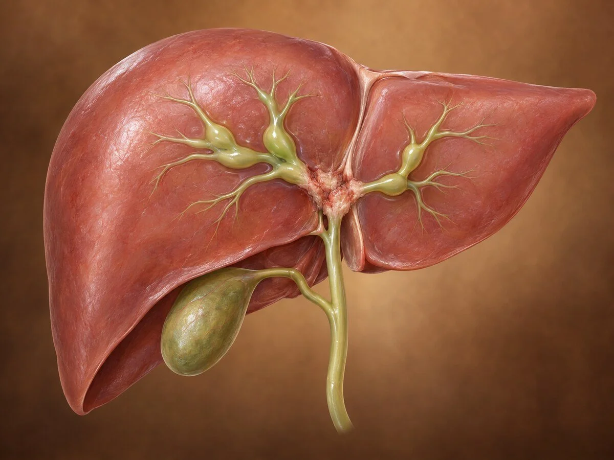
Cholangiocarcinoma is cancer arising from the lining of the bile ducts. It is not one disease but three, classified by where along the biliary tree it arises — and the distinction matters more than almost anything else, because each requires a completely different operation and carries a different outlook.
The three types
Intrahepatic — within the liver, from the small ducts. Presents as a liver mass; treated by liver resection. Covered in detail on its own page under Liver.
Perihilar (Klatskin tumour) — at the junction where the right and left hepatic ducts meet. The commonest type, accounting for roughly half to two-thirds of cases, and the most technically demanding to treat.
Distal — the lower bile duct, below the cystic duct. Treated by pancreaticoduodenectomy, and discussed alongside the periampullary tumours.
This page concentrates on the perihilar form, which is where the surgical challenge is greatest.
How it forms
Cholangiocytes lining the bile ducts are injured by chronic inflammation, and repeated cycles of damage and repair accumulate genetic errors until cancer develops. The recognised causes all share that theme:
Primary sclerosing cholangitis — the strongest identified risk factor, carrying a substantial lifetime risk.
Choledochal cysts and Caroli disease — congenital dilatations of the biliary tree, with a risk that persists and is one of the main reasons these cysts are excised rather than drained.
Hepatolithiasis — stones within the intrahepatic ducts.
Liver fluke infection — a major cause in parts of Southeast Asia, rare here.
Chronic viral hepatitis and cirrhosis, and certain chemical exposures.
In a large proportion no risk factor is identified. Patients frequently search for something they did wrong, and often there is nothing.
The tumour typically grows along the duct wall rather than as a discrete ball — a periductal-infiltrating pattern — extending longitudinally beneath an intact lining. This explains two clinically important things: the tumour is often much more extensive than it appears on imaging, and achieving a clear margin requires removing considerably more duct than the visible abnormality suggests.
Symptoms
Painless obstructive jaundice — the typical presentation
itching, often severe
dark urine and pale stools
weight loss and anorexia
vague upper abdominal discomfort
cholangitis, particularly after biliary instrumentation
How the diagnosis is made
MRI with MRCP is the central investigation, defining the extent of ductal involvement — how far the tumour reaches into the right and left ductal systems, which determines resectability. Contrast CT assesses vascular involvement of the portal vein and hepatic artery, and looks for nodal and distant disease. Volumetry measures the future liver remnant.
Tissue diagnosis can be difficult. Brushings obtained at ERCP have limited sensitivity, and a negative result does not exclude cancer. Where imaging is characteristic and the disease appears resectable, surgery may reasonably proceed without histological confirmation — one of the few situations in oncology where that is accepted practice.
CA 19-9 is usually raised but is unreliable in the presence of jaundice.
The Bismuth–Corlette classification describes how far the tumour extends into the hepatic ducts and guides the operation required.
A word of caution about stenting: placing stents before a surgical assessment can make subsequent imaging harder to interpret and introduces infection. Where resection is contemplated, patients are best assessed at a specialist unit before the biliary tree is instrumented.
Treatment
Surgery
Complete resection with clear margins is the only curative option, and for perihilar tumours it is major surgery. The standard operation combines:
excision of the extrahepatic bile duct
major liver resection — usually right or left hepatectomy, frequently extended
caudate lobe resection, because the caudate ducts drain directly into the hilum and are involved early; omitting it compromises the margin
regional lymphadenectomy
biliary reconstruction, joining the remaining ducts to a loop of small bowel
portal vein resection and reconstruction where required
Because a major liver resection is required in an often jaundiced patient, preparation matters: biliary drainage of the future liver remnant, and portal vein embolisation to induce growth of the side that will remain. These preparatory steps take weeks and are part of why this disease belongs in a specialist unit from the outset.
Liver transplantation, combined with neoadjuvant chemoradiotherapy and under strict protocol, has a role in highly selected patients with early unresectable perihilar tumours, particularly those arising in primary sclerosing cholangitis.
Chemotherapy and targeted treatment
Adjuvant chemotherapy follows resection. For unresectable or metastatic disease, chemotherapy combined with immunotherapy is the current standard. Molecular profiling is routine, as FGFR2 fusions and IDH1 mutations occur in a proportion and have specific licensed treatments — a genuine change in the outlook for advanced disease over the past few years.
Palliative biliary drainage
Where resection is not possible, relieving jaundice by endoscopic or percutaneous stenting improves symptoms, particularly itching, and allows chemotherapy to proceed. Photodynamic therapy and radiofrequency ablation within the duct have selective roles.
Recovery
Major hepatectomy with biliary reconstruction is substantial surgery, with a hospital stay typically of ten days to two weeks and recovery over two to three months. Bile leak and liver insufficiency are the specific concerns, and this is reflected in outcomes being clearly better in high-volume centres.
Follow-up
Regular imaging, liver function tests and CA 19-9. Patients with a biliary-enteric anastomosis remain at risk of cholangitis lifelong and should be advised to seek attention promptly with fever and rigors.
Common questions
Is bile duct cancer curable?
It can be, when the tumour can be completely removed. That depends chiefly on how far it extends along the ducts and whether the major blood vessels are involved, rather than on its size. Even when cure is not possible, treatment has improved considerably in recent years and can control the disease for a substantial period.
Why do I need part of my liver removed for a bile duct cancer?
Because the tumour spreads along the duct wall, often further than it appears on scans. Removing the duct alone would leave cancer behind. Taking the liver on one side, together with the ducts, is what makes a clear margin achievable.
What is a Klatskin tumour?
The name given to a cholangiocarcinoma at the point where the right and left bile ducts join, just outside the liver. It is the commonest location and the most complex to operate on because of its position among the major blood vessels.
I have been told it is inoperable — is that final?
Not necessarily. Whether a perihilar tumour can be removed depends heavily on the techniques available to the assessing team — portal vein embolisation to grow the remaining liver, vascular reconstruction, and experience with extended resections. A second opinion at a specialist unit is reasonable before accepting that surgery is impossible.
Should I have a stent put in before seeing a surgeon?
If you have infection or severe symptoms, drainage may be urgent. Otherwise, where surgery is being considered, it is often better to be assessed at a specialist centre first, because stents can make the scans harder to interpret and introduce infection. This is worth asking about specifically.
Did something cause this?
In most patients, no identifiable cause is found. Certain conditions increase the risk — primary sclerosing cholangitis, congenital bile duct cysts, long-standing duct stones — but the majority of people who develop it have none of these.
Related conditions
Other conditions of the gallbladder & bile ducts covered on this site:
This page provides general information and does not replace an individual medical consultation. Assessment and treatment are decided for each patient after review of their history, examination and imaging, and in the setting of a multidisciplinary team.
