

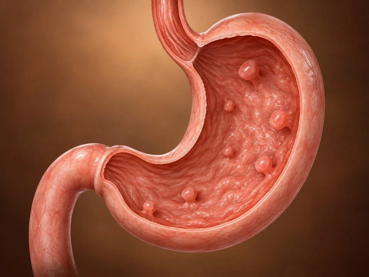
Gastric polyps are growths projecting from the stomach lining. Most are found incidentally during an endoscopy performed for something else, and most are entirely harmless. Their importance lies almost entirely in accurate classification, because the different types have completely different implications — from requiring nothing at all, to indicating an inherited cancer syndrome. The single most useful action when a polyp is found is a biopsy, and of the surrounding stomach lining as well as of the polyp itself.
The types, and how they form
Fundic gland polyps
Now the commonest type in Western practice. They are small, multiple, glassy-looking polyps in the body and fundus, formed by cystic dilatation of the gastric glands.
They arise in two quite different settings:
Sporadic, associated with proton pump inhibitor use. Prolonged acid suppression raises gastrin, which has a trophic effect on the parietal cells; these become hypertrophied and obstruct the gland necks, and the glands behind them dilate into cysts. These polyps carry essentially no malignant potential and frequently regress if the medication is stopped. They are not a reason to stop a proton pump inhibitor that is genuinely needed.
Syndromic, in familial adenomatous polyposis. Here fundic gland polyps are typically numerous — often dozens or hundreds — appear in younger patients, and may contain dysplasia. Numerous fundic gland polyps in a young patient not taking acid suppression should prompt consideration of FAP and assessment of the colon. This is the clinically important distinction.
Hyperplastic polyps
These are a regenerative response to chronic injury. Persistent inflammation — from Helicobacter pylori gastritis, autoimmune atrophic gastritis, bile reflux, or around an ulcer or surgical anastomosis — drives exaggerated repair, producing elongated, distorted, branching foveolar glands with inflamed stroma.
Two consequences follow. First, larger hyperplastic polyps, particularly those above 1cm, may contain dysplasia or focal carcinoma. Second, and more important, the polyp is a marker of the underlying gastritis, and it is the inflamed stomach around the polyp, not the polyp itself, that carries the greater cancer risk. Hence the rule: biopsy the surrounding mucosa, treat the cause, and many of these polyps regress after H. pylori eradication.
Gastric adenomas (intestinal-type dysplastic polyps)
These are true neoplastic polyps — dysplastic by definition — and are the direct precursors of gastric adenocarcinoma, following the same sequence as adenomas in the colon. They arise in stomachs with atrophic gastritis and intestinal metaplasia, usually in the antrum. Risk of harbouring carcinoma rises with size, villous architecture and high-grade dysplasia.
All adenomas should be removed completely, and the surrounding stomach mapped by biopsy, because the field from which they arose remains at risk.
Other types
Inflammatory fibroid polyp — a benign submucosal lesion of spindle cells and eosinophils, harmless, occasionally causing obstruction or intussusception if large.
Hamartomatous polyps — occurring in Peutz-Jeghers syndrome, juvenile polyposis and Cowden syndrome, each with their own surveillance requirements.
Neuroendocrine tumours (gastric carcinoids) — may appear as small polyps and are covered on their own page.
Gastrointestinal stromal tumours — submucosal rather than mucosal, appearing as a smooth bulge with normal overlying lining, and covered separately.
Xanthoma — a small yellow plaque of lipid-laden macrophages, benign, but associated with atrophic gastritis and therefore a reason to assess the background mucosa.
Symptoms
Almost always none. Larger polyps may cause:
iron-deficiency anaemia from chronic slow bleeding
dyspepsia or vague upper abdominal discomfort
obstruction, if a large antral polyp prolapses into the pylorus — producing intermittent vomiting
rarely, overt bleeding
Symptoms attributed to a small polyp are usually caused by something else, and it is worth saying so rather than removing a polyp and leaving a patient with unchanged symptoms and a false sense of resolution.
How the diagnosis is made
Endoscopy identifies the polyps, and histology classifies them. A complete assessment includes:
the number, size, location and appearance of the polyps
biopsy or removal of the polyp — the largest ones, and a representative sample where there are many
biopsies of the non-polypoid stomach lining, from antrum and body, to establish whether there is H. pylori gastritis, atrophy or intestinal metaplasia. This step is the one most often skipped and the one that most often changes management.
testing for H. pylori
assessment of the duodenum, and consideration of colonoscopy where fundic gland polyps are numerous or hamartomatous polyps are found
Treatment
Small sporadic fundic gland polyps — sample one or two to confirm the diagnosis; the rest need nothing. No surveillance is required. Review of the need for ongoing acid suppression is reasonable, but the polyps themselves are not a reason to stop necessary treatment.
Hyperplastic polyps — remove those above about 1cm, those that are symptomatic, and any with concerning features. Eradicate H. pylori and re-endoscope after some months, as many of the remainder disappear.
Adenomas — remove completely, by snare polypectomy, endoscopic mucosal resection or endoscopic submucosal dissection according to size and appearance. Confirm complete excision histologically.
Polyps with high-grade dysplasia or early carcinoma — endoscopic submucosal dissection gives an intact specimen and accurate assessment of depth and margins, and is curative for lesions meeting defined criteria. Surgery is reserved for lesions beyond those criteria.
Very numerous polyps, or polyps in a recognised syndrome — management follows the syndrome's protocol, and genetic assessment is arranged.
Surgery for gastric polyps as such is now rare, and is reserved for large lesions unsuitable for endoscopic removal, for bleeding or obstruction not otherwise controllable, and for polyps proving to be carcinoma.
Follow-up
Follow-up is determined by the histology and the state of the background mucosa, not by the fact that a polyp was found.
sporadic fundic gland polyps — none
hyperplastic polyps with treated gastritis — a check endoscopy, then discharge if clear
adenoma, or atrophic gastritis with extensive intestinal metaplasia — endoscopic surveillance at intervals, since the whole mucosa carries risk
dysplasia — closer surveillance, with mapped biopsies
polyposis syndromes — lifelong protocol-driven surveillance of stomach, duodenum and colon
When to seek an opinion
Iron-deficiency anaemia without obvious cause, persistent indigestion with alarm features, or a known diagnosis of gastric polyps without a clear explanation of what type they are and what follow-up is needed. A family history of gastric or bowel cancer, or of polyposis, is a reason to seek advice specifically about screening.
Common questions
I was told I have polyps in my stomach. Is that cancer?
No. A polyp is a small growth of the lining, and the great majority are benign. What matters is the type, which is determined by examining a sample under the microscope. Some types carry no risk at all, while a minority need removing because they can change over time.
Are they caused by my acid tablets?
One common type — small, multiple polyps in the upper part of the stomach — is associated with taking acid-suppressing tablets for a long time. These carry essentially no risk of becoming cancer, and they often disappear if the tablets are stopped. They are not in themselves a reason to stop a medication you need.
Do they all have to be removed?
No. Small harmless ones are usually sampled to confirm the type and then left alone. Ones that are larger, or of a type that can change, are removed through the endoscope, which is a straightforward procedure with no incisions.
Why were biopsies taken from the normal-looking stomach as well?
Because in several cases the polyp is a sign rather than the problem. Long-standing inflammation of the stomach lining, often from a bacterial infection, produces polyps and also carries its own small risk. Sampling the surrounding lining tells us whether that inflammation is present and needs treating, which is frequently the more important finding.
Will they come back?
Some types do, particularly if the underlying cause persists. Treating the bacterial infection often makes them disappear and stops new ones forming. Whether any further check is needed depends on the type found, and this should be stated clearly to you rather than left vague.
Should my family be checked?
Usually not. The exception is where the polyps are very numerous, or of a particular inherited type, which can point to a condition that runs in families and also affects the bowel. In that situation genetic assessment and checks for relatives are recommended, and this is one of the reasons the type of polyp matters so much.
Do I need a repeat endoscopy?
It depends entirely on what the samples showed. Some people need none at all; others need a check after treatment of an infection; a minority with changes in the lining need regular surveillance. You should be told which group you are in, and why.
What symptoms do stomach polyps cause?
Almost always none; most are found by chance at gastroscopy. Larger polyps can cause iron-deficiency anaemia from slow bleeding, indigestion or upper abdominal discomfort, intermittent vomiting if a polyp near the outlet of the stomach blocks it, and rarely visible bleeding. Symptoms blamed on a small polyp usually have another cause.
How are stomach polyps treated?
It depends on the type. Small fundic gland polyps need nothing beyond confirming the diagnosis. Larger hyperplastic polyps are removed and H. pylori, if present, is treated. Adenomas are removed completely at endoscopy. Surgery is now rarely needed and is reserved for polyps that cannot be removed endoscopically, bleeding or obstruction that cannot otherwise be controlled, or cancer.
Related conditions
Other conditions of the stomach covered on this site:
This page provides general information and does not replace an individual medical consultation. Assessment and treatment are decided for each patient after review of their history, examination and investigations.
