

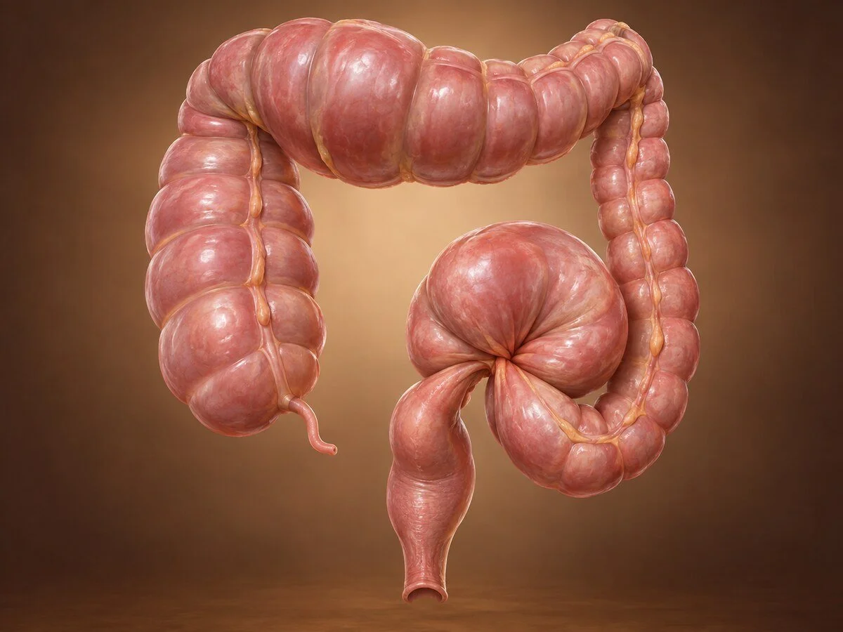
Large bowel obstruction is a surgical emergency. Unlike small bowel obstruction, which is usually caused by adhesions and often settles without an operation, obstruction of the colon is most often caused by a cancer and rarely resolves on its own. It also carries a particular danger: in a proportion of patients the valve between small and large bowel prevents any decompression backwards, and the colon becomes a closed loop that distends until it perforates. Recognising that situation is the most urgent part of the assessment.
How it develops
The colon can be blocked by something growing within it, by the bowel twisting on itself, or by the muscle simply ceasing to work.
Mechanical causes
Colorectal cancer — the commonest cause in adults, accounting for the majority. Typically in the left colon, where the lumen is narrower and the stool formed.
Volvulus — a twist of the bowel on its mesentery, discussed below.
Diverticular stricture — from repeated inflammation and fibrosis.
Hernia containing colon, adhesions (much less common in the colon than the small bowel), intussusception (in adults, usually with a tumour as the lead point), faecal impaction, and extrinsic compression by pelvic or other tumours.
Anastomotic stricture after previous surgery; ischaemic stricture after a previous episode of colonic ischaemia.
The closed loop — why some patients deteriorate fast
This is the point that determines urgency. If the ileocaecal valve is competent — which it is in a substantial proportion of people — gas and fluid entering the colon from above cannot escape backwards into the small bowel, and cannot pass the obstruction ahead. The colon becomes a closed loop.
Pressure rises steadily, and by Laplace's law, wall tension is greatest where the radius is greatest — the caecum. So although the obstruction may be in the sigmoid, it is the caecum that becomes ischaemic and perforates. A caecal diameter beyond about 10–12cm on plain film is the accepted warning threshold, and a tender right iliac fossa in a patient with left-sided obstruction is an ominous clinical sign.
Conversely, an incompetent valve allows the small bowel to decompress the colon, which produces a more gradual picture with vomiting and a lower risk of perforation.
Volvulus
Volvulus requires two conditions: a segment of bowel on a long mesentery, and a narrow base about which it can rotate.
Sigmoid volvulus — much the commoner. Chronic constipation and a high-residue diet produce a long, redundant, heavy sigmoid loop; repeated episodes cause the mesentery to shorten and its base to narrow, which makes further twisting easier. It is commonest in elderly, institutionalised and neurologically impaired patients, and in some geographical regions — the "volvulus belt" of Africa, the Middle East and parts of Asia — it is far commoner than in Western practice. Rotation is usually anticlockwise; a twist beyond 180° obstructs the lumen, and beyond 360° obstructs the blood supply.
Caecal volvulus — requires a congenital failure of fixation of the right colon to the posterior abdominal wall, present in a minority of people. It occurs in younger patients than sigmoid volvulus, and a variant, caecal bascule, involves the caecum folding upward without rotation.
Transverse colon volvulus — rare.
Pseudo-obstruction (Ogilvie's syndrome)
Massive colonic dilatation with no mechanical obstruction, caused by an imbalance of autonomic supply to the colon — typically excess sympathetic or deficient parasympathetic activity. It occurs in unwell, immobile hospital inpatients: after orthopaedic surgery, with severe infection, cardiac or respiratory failure, electrolyte disturbance, or on opioids and anticholinergic drugs. It matters enormously that it is distinguished from mechanical obstruction, because the treatment is entirely different and operating on it is harmful.
Symptoms
Absolute constipation — no stool and no flatus passed
Abdominal distension, often marked, and characteristically more pronounced than in small bowel obstruction
Colicky abdominal pain, which becomes constant and severe if the bowel becomes ischaemic
Vomiting — late, and faeculent in advanced cases; may be absent entirely with a competent ileocaecal valve
a history suggesting the cause: altered bowel habit, rectal bleeding and weight loss over preceding months point to cancer; previous similar self-resolving episodes point to volvulus
Warning features of impending or actual perforation: localised tenderness, particularly in the right iliac fossa; guarding and rebound; fever; tachycardia; hypotension; and a rising lactate. These convert an urgent situation into an immediate one.
How the diagnosis is made
CT of the abdomen and pelvis with contrast — the investigation of choice. It confirms obstruction, identifies the level and cause, measures caecal diameter, detects free gas and signs of ischaemia, and stages a tumour.
Plain abdominal X-ray — may show dilated colon with the peripheral haustral pattern; in sigmoid volvulus, the classical "coffee bean" or "bent inner tube" sign of a hugely dilated loop arising from the pelvis toward the right upper quadrant.
Water-soluble contrast enema — distinguishes mechanical obstruction from pseudo-obstruction where CT is equivocal.
Flexible sigmoidoscopy — both diagnostic and therapeutic in sigmoid volvulus.
Blood tests including lactate, and correction of electrolytes.
Treatment
Initial management in all cases: nil by mouth, intravenous fluids, nasogastric decompression if vomiting, correction of electrolytes, analgesia, catheterisation, and antibiotics where perforation or ischaemia is suspected.
Obstructing colorectal cancer
Right-sided obstruction — right hemicolectomy with primary anastomosis is usually safe and is the standard approach, since the small bowel tolerates a join well.
Left-sided obstruction — the choice is genuinely nuanced:
Self-expanding metal stent as a bridge to surgery — placed endoscopically to relieve the obstruction, allowing the patient to be resuscitated, staged properly, and operated on electively and laparoscopically days or weeks later, frequently avoiding a stoma altogether. This has transformed management for suitable patients. It is also the treatment of choice for palliation where the disease is incurable. Perforation is the principal risk, and stenting is avoided where perforation has occurred or the tumour is very proximal.
Hartmann's procedure — resection with end colostomy. Safe in the unstable or contaminated patient, but a considerable proportion of stomas are never reversed.
Resection with primary anastomosis, with or without on-table lavage and a protecting ileostomy — appropriate in stable patients with healthy bowel.
Subtotal colectomy — where the caecum is compromised or synchronous tumours are present.
Sigmoid volvulus
Endoscopic decompression by flexible sigmoidoscopy, with a flatus tube left in place, is the initial treatment and succeeds in the great majority. It is dramatic and immediately relieving. It is contraindicated where there are signs of ischaemia or peritonitis, and mucosal ischaemia seen at endoscopy mandates surgery.
Recurrence after decompression alone is high — the majority within two years — so elective sigmoid colectomy during the same admission is recommended for patients fit for it. Repeated decompression without definitive surgery eventually results in an emergency presentation with gangrene, which carries a far higher mortality.
Emergency surgery is required for failed decompression or ischaemia: sigmoid resection, with primary anastomosis in stable patients or Hartmann's procedure otherwise.
Caecal volvulus
Endoscopic reduction rarely succeeds. Treatment is surgical: right hemicolectomy with ileocolic anastomosis is the standard and definitive option. Caecopexy has a higher recurrence rate and is reserved for patients unfit for resection.
Pseudo-obstruction
Treatment is medical and supportive: correct electrolytes (particularly potassium, magnesium and calcium), stop opioids and anticholinergics, treat the underlying illness, mobilise the patient, and decompress with a nasogastric tube and flatus tube. Neostigmine is highly effective and often produces dramatic decompression, given with cardiac monitoring because of the risk of bradycardia. Colonoscopic decompression is used where this fails. Surgery is a last resort for perforation or ischaemia and carries high mortality.
Recovery
Recovery depends on the operation and on whether the presentation was with perforation or ischaemia. Elective surgery after stenting follows the usual laparoscopic colorectal course of three to five days. Emergency resection is a longer recovery, often with a stoma and frequently with a period in critical care. Stoma reversal, where planned, is usually considered after three to six months.
When to seek help urgently
Not passing stool or wind, with a distended painful abdomen, requires emergency assessment. Increasing pain, fever or feeling very unwell in that setting requires it immediately.
Common questions
Why is a blocked large bowel more urgent than a blocked small bowel?
Because it rarely settles by itself, it is usually caused by something that needs treating, and in many people the valve between small and large bowel prevents the pressure escaping backwards. The bowel then keeps distending until it is at risk of bursting, usually at the far end from the blockage.
Does a blockage always mean cancer?
No, but it is the commonest cause in adults and must be excluded. Other causes include a twist of the bowel, scarring from diverticular inflammation, and — in unwell hospital patients — a condition where the bowel simply stops working without any physical blockage at all.
What is a stent and would it help me?
A stent is an expanding metal tube placed through a colonoscope to open the blocked segment. It relieves the blockage within a day or so, which allows you to recover, be properly scanned and then have a planned keyhole operation instead of an emergency one — often avoiding a stoma entirely. It is not suitable for every situation, particularly where the bowel has already perforated.
Will I need a stoma?
Not always. Emergency operations more often require one than planned operations, which is part of the reason relieving the blockage first with a stent, where possible, is valuable. Many stomas formed in an emergency are temporary and can be reversed after a few months, though this requires a second operation and not everyone chooses to have it.
The twist was untwisted with a camera. Why do I need an operation as well?
Because untwisting relieves the emergency but does not change the anatomy that allowed it. Most people whose bowel has twisted once will have it happen again, often within a couple of years, and the second episode may cut off the blood supply, which is far more dangerous. Removing the redundant loop during the same admission, if you are fit enough, prevents that.
My scan shows a hugely swollen bowel but no blockage. What does that mean?
This is a condition in which the nerves controlling the bowel muscle become unbalanced during serious illness, and the bowel dilates without anything physically obstructing it. It is treated by correcting the salts in the blood, stopping certain painkillers, getting you moving, and sometimes a specific drug that restores bowel activity. Operating on it would be harmful, which is why distinguishing it from a true blockage matters so much.
How soon must this be treated?
Same day. A blocked large bowel is treated as an emergency, and increasing pain, fever or tenderness over the right side of the abdomen means it needs attention immediately rather than within hours.
Related conditions
Other conditions of the colon & rectum covered on this site:
This page provides general information and does not replace an individual medical consultation. Large bowel obstruction is a medical emergency requiring immediate hospital assessment.
