

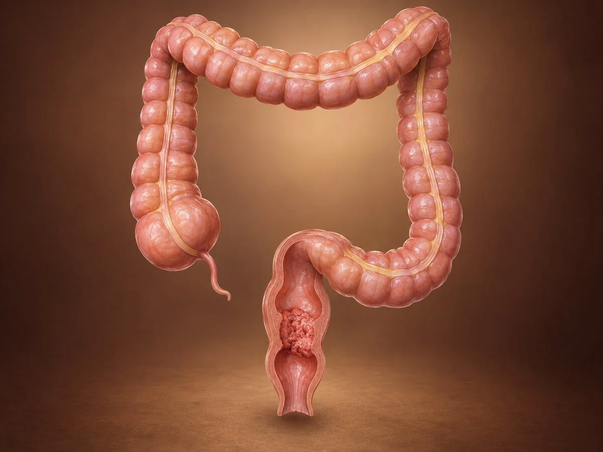
Rectal cancer arises in the last fifteen centimetres of the large bowel. Although it shares its biology with colon cancer, it is managed quite differently, and the reason is anatomical: the rectum lies deep in the bony pelvis, surrounded by structures that cannot be sacrificed, with no serosal covering over much of its circumference and immediately adjacent to the sphincters that preserve continence. Everything that distinguishes rectal from colon cancer — the use of radiotherapy, the meticulous surgical technique, the emphasis on MRI before treatment, the question of whether surgery can be avoided altogether — follows from that confined anatomy.
How it forms
The underlying process is the same adenoma-carcinoma sequence described for colon cancer: progressive genetic damage — APC, KRAS, SMAD4, TP53 — transforming a benign polyp into an invasive tumour over ten to fifteen years.
What differs is what happens once the tumour invades outward. The rectum is enclosed in the mesorectum, a fatty envelope containing its blood vessels and lymph nodes, bounded by a thin but definite layer, the mesorectal fascia. Tumour spreads first within this envelope. The fascia acts as a natural barrier, and the distance from the tumour to it — the circumferential resection margin — is the single most important predictor of local recurrence.
This is the insight behind total mesorectal excision: removing the rectum together with its entire mesorectal envelope intact, dissecting in the areolar plane immediately outside the fascia rather than cutting into the fat. When this is done well, local recurrence falls from a historical figure of around a quarter of patients to a small single-figure percentage. Few technical changes in surgery have had so large an effect, and it explains why rectal cancer surgery is concentrated in specialist units.
The rectum also drains partly to lateral pelvic nodes and, through the lower rectal veins, into the systemic circulation, which is why lung metastases are relatively more common in rectal than in colon cancer.
Symptoms
Rectal bleeding — the commonest symptom, typically fresh blood on or mixed with the stool
Change in bowel habit — looser, more frequent stools
Tenesmus — a persistent sensation of incomplete emptying, of needing to go again immediately afterwards. This is characteristic of a rectal tumour and is often described by patients as feeling that "something is still there".
mucus discharge
narrowing of stool calibre
pelvic or perineal pain, or pain on sitting — a later feature suggesting local invasion
anaemia, weight loss
incontinence or a change in continence
Rectal bleeding should not be attributed to haemorrhoids without examination of the rectum. A digital rectal examination detects a substantial proportion of rectal cancers and takes a moment; omitting it is the commonest reason for delay.
How the diagnosis is made
Accurate staging before treatment matters more in rectal cancer than almost anywhere else, because it determines whether radiotherapy is given, and radiotherapy cannot be undone.
Digital rectal examination — assesses the distance from the anal verge, mobility, and sphincter involvement.
Rigid or flexible sigmoidoscopy with biopsy — establishes the diagnosis and the exact height of the tumour, which determines the operation.
Full colonoscopy — to exclude synchronous tumours elsewhere in the colon.
High-resolution pelvic MRI — the key investigation. It shows depth of invasion, the relationship to the mesorectal fascia, nodal involvement, extramural venous invasion, and involvement of adjacent organs or the sphincter complex. It is what decides the treatment plan.
Endorectal ultrasound — more accurate than MRI for very early tumours, where the decision is between local excision and radical surgery.
CT of chest, abdomen and pelvis — for distant disease.
CEA, and mismatch repair testing on all tumours.
Treatment
Treatment is planned by a specialist multidisciplinary team and depends on the height of the tumour, its stage on MRI, and the patient's continence and fitness.
Very early tumours
Small, superficial, well-differentiated tumours without adverse features can be removed by transanal endoscopic microsurgery or transanal minimally invasive surgery — full-thickness local excision through the anus, with no abdominal incision and no stoma. Pathology of the specimen then determines whether radical surgery is needed.
Neoadjuvant treatment
Given before surgery for locally advanced tumours — those threatening or involving the mesorectal fascia, with significant nodal disease, or with extramural venous invasion.
Long-course chemoradiotherapy — five weeks of radiotherapy with chemotherapy, followed by surgery after an interval. Shrinks the tumour, increases the chance of a clear margin, and reduces local recurrence.
Short-course radiotherapy — five days, followed by early or delayed surgery.
Total neoadjuvant therapy — giving all the chemotherapy as well as the radiotherapy before surgery. This has become increasingly standard: it improves completion rates, increases complete response, and treats micrometastatic disease early.
Immunotherapy alone for mismatch repair-deficient rectal cancers, a small group in whom results have been remarkable, with complete clinical response in a high proportion and the prospect of avoiding both radiotherapy and surgery.
Watch and wait
Some tumours disappear completely on clinical, endoscopic and MRI assessment after neoadjuvant treatment — a complete clinical response. In these patients, intensive surveillance rather than immediate surgery is a recognised option, preserving the rectum and avoiding a permanent stoma. It requires a committed programme of frequent endoscopy and MRI, because regrowth occurs in a minority and is almost always salvageable if detected promptly. It is a genuine choice, with a trade-off, and it should be offered and explained rather than assumed either way.
Surgery
Anterior resection with total mesorectal excision — the standard operation, removing the rectum and its mesorectum and rejoining the colon to the remaining rectum or anus. A temporary ileostomy is commonly formed to protect a low join, and reversed after some months.
Abdominoperineal excision — for tumours involving the sphincters or so low that a join is impossible. The rectum and anus are removed and a permanent colostomy is formed. This is a good operation performed for the right reasons, and patients live full lives with a stoma; it is not a failure of treatment.
Intersphincteric resection — preserves continence in selected very low tumours.
Pelvic exenteration — for locally advanced tumours invading adjacent organs, in specialist centres.
Laparoscopic, robotic and transanal approaches are all used. Robotic surgery has particular advantages in the narrow male pelvis. Careful nerve preservation during the dissection protects bladder and sexual function, and this is an explicit objective of the operation, not an incidental one.
After surgery
Adjuvant chemotherapy is given according to the final pathology and what was given beforehand.
Recovery and function
Hospital stay after anterior resection is typically five to eight days. Return to normal activity takes six to eight weeks.
Patients deserve a frank account of function, because this is where expectations are most often mismatched:
Low anterior resection syndrome is common after removal of the rectum: frequency, urgency, fragmentation of stool, clustering of bowel motions, and variable incontinence. It improves over one to two years but may not disappear. It is managed with diet, fibre and antidiarrhoeal agents, pelvic floor physiotherapy, transanal irrigation and, occasionally, sacral nerve stimulation. Knowing about it in advance makes it considerably easier to live with.
Bladder and sexual dysfunction can follow pelvic nerve injury, and radiotherapy contributes. This should be discussed before treatment, including fertility preservation in younger patients, since radiotherapy to the pelvis affects fertility in both sexes.
Stoma — temporary or permanent, with specialist stoma nurse support, which makes a substantial difference to adjustment.
Follow-up
Clinical review, CEA, CT at intervals, and colonoscopy. Patients on a watch-and-wait programme require much more intensive surveillance, with endoscopy and MRI every few months in the first two years.
When to seek an opinion
Rectal bleeding persisting more than a few weeks at any age; a change in bowel habit; tenesmus or a persistent feeling of incomplete emptying; mucus discharge; or unexplained anaemia. Rectal cancer treatment should be planned by a multidisciplinary team with specialist MRI reporting, and the surgery performed by a surgeon who does it regularly.
Common questions
Will I definitely need a permanent bag?
No — most people do not. Whether a permanent stoma is needed depends chiefly on how close the tumour is to the muscles that control continence. Most rectal cancers can be removed with the bowel rejoined. A temporary stoma to protect the join for a few months is common, and is reversed afterwards.
Why do I need radiotherapy when my friend with bowel cancer did not?
Because the rectum sits in a confined bony pelvis where there is little room to take a wide margin, so treatment given beforehand to shrink the tumour reduces the chance of it returning locally. The colon lies in the open abdomen, where an adequate margin is easier to achieve, so radiotherapy is not normally used.
I have been told the tumour disappeared after treatment. Do I still need surgery?
Not necessarily. When the tumour has apparently disappeared completely on examination, endoscopy and scanning, careful monitoring instead of surgery is a recognised option that preserves the rectum. It requires frequent checks, because in a minority the tumour regrows — almost always treatable if caught early. It is a genuine choice, and worth discussing in detail.
Will my bowels work normally afterwards?
Often differently. Because the rectum acts as a reservoir, removing it commonly causes frequency, urgency and stools broken into several visits, particularly in the first year. This usually improves substantially over one to two years, and there is a good deal that helps — diet, medication, pelvic floor physiotherapy and other treatments. It is better to expect this than to be surprised by it.
Will it affect sex or fertility?
It can. The nerves controlling sexual function run close to the rectum and are carefully preserved during surgery, but function may still change, and radiotherapy to the pelvis affects fertility in both men and women. This should be discussed before treatment starts, and sperm or egg storage arranged where appropriate. It is a reasonable thing to raise, and a good team will raise it first.
Why is MRI so important?
Because it shows how close the tumour is to the envelope of tissue around the rectum, which determines whether treatment is needed before surgery and how the operation is planned. It is the single test that most influences the treatment decision in rectal cancer.
Does it matter where I have the surgery?
Yes, more than for most operations. The technique of removing the rectum within its intact envelope is what determines whether the cancer returns locally, and results are consistently better in units and with surgeons who perform this regularly. It is entirely reasonable to ask.
Can rectal cancer be cured?
Yes, frequently. Early tumours are cured in the great majority of cases, and even locally advanced tumours are often cured with combined treatment. Outlook depends on stage and on the quality of the surgery, which is why the staging and the choice of unit both matter.
Related conditions
Other conditions of the colon & rectum covered on this site:
This page provides general information and does not replace an individual medical consultation. Assessment and treatment are decided for each patient after review of their history, examination, investigations and discussion in a multidisciplinary meeting.
