

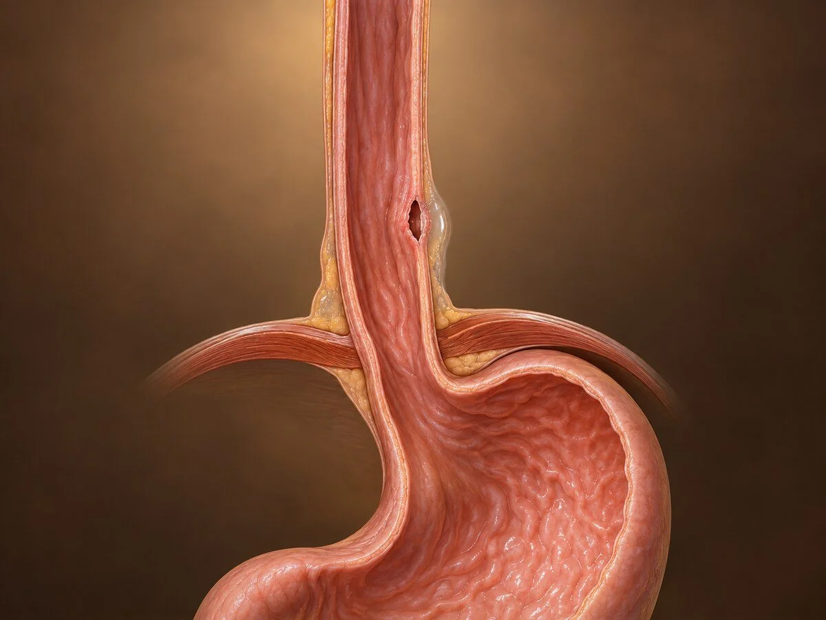
Perforation of the oesophagus is one of the true emergencies of general surgery. It is uncommon, it is frequently mistaken for a heart attack or a chest infection in its first hours, and the delay that follows that mistake is the single greatest determinant of whether the patient survives. Treated within the first twelve to twenty-four hours the outlook is good; treated after seventy-two hours it becomes considerably worse. Everything on this page follows from that one fact.
How it happens
The oesophagus is uniquely vulnerable among hollow organs, and for anatomical reasons worth stating plainly:
it has no serosa — no outer containing layer — so a tear is not walled off as it would be elsewhere in the gut
it lies in the mediastinum, a space of loose areolar tissue with no barriers, so contamination spreads rapidly upward and downward
negative intrathoracic pressure during inspiration actively draws saliva, gastric contents and swallowed air into the mediastinum through the tear
the contamination is chemical as well as bacterial — gastric acid and digestive enzymes cause immediate necrotising mediastinitis, not merely infection
The result is a fulminant chemical and bacterial mediastinitis, followed within hours by empyema and systemic sepsis.
Causes
Iatrogenic perforation is the commonest cause, accounting for the majority. It follows endoscopy — particularly therapeutic procedures such as dilatation of a stricture, achalasia balloon dilatation, stent insertion, endoscopic resection or variceal treatment — and also occurs with nasogastric tube placement, transoesophageal echocardiography, and during surgery on adjacent structures. The risk is concentrated at the natural narrowings: the cricopharyngeus, the level of the aortic arch and left main bronchus, and the hiatus.
Boerhaave syndrome is spontaneous rupture caused by a sudden rise in intraluminal pressure against a closed cricopharyngeus — classically forceful vomiting or retching after a large meal and alcohol, but also described after weightlifting, childbirth, coughing and defecation. The tear is characteristically a longitudinal split in the left posterolateral wall of the lower oesophagus, just above the diaphragm, where the muscle layer is thinnest. It is the most lethal form, because the tear is full-thickness, the contamination is gastric, and the diagnosis is the one most often missed.
Other causes: foreign body ingestion, particularly sharp objects or impacted food boluses and disc batteries in children; caustic ingestion; penetrating or blunt trauma; and perforation through a tumour or through an existing deep ulcer.
A Mallory-Weiss tear is a related but distinct entity: a mucosal tear only, in the same setting of retching, causing bleeding rather than perforation, and usually settling without intervention. Distinguishing the two is a matter of whether the tear is partial or full thickness.
Symptoms
The classical description for spontaneous rupture is Mackler's triad: vomiting, severe chest pain, and subcutaneous emphysema. It is present in a minority of patients, and waiting for it causes delay.
Severe, sudden chest pain, often radiating to the back or the left shoulder, typically beginning immediately after vomiting or after an endoscopic procedure
neck or epigastric pain, depending on the level of the tear
pain on swallowing, and refusal to swallow
breathlessness
surgical emphysema — crackling under the skin of the neck or chest wall, a specific but late sign
fever, tachycardia, hypotension — sepsis developing over hours
Hamman's sign — a crunching sound in time with the heartbeat, from mediastinal air
The pattern that should trigger suspicion: chest pain after vomiting, or chest, neck or abdominal pain after any endoscopic procedure, is oesophageal perforation until proved otherwise. It is commonly misdiagnosed initially as myocardial infarction, pulmonary embolism, perforated peptic ulcer, pancreatitis or pneumonia, and the diagnosis is often made only when the patient fails to respond to treatment for one of these.
How the diagnosis is made
Chest X-ray may show mediastinal air, surgical emphysema, pleural effusion (typically left-sided), pneumothorax or hydropneumothorax. It can be normal in the first hours, and a normal film does not exclude the diagnosis.
CT with oral water-soluble contrast is the investigation of choice. It demonstrates the leak, locates it, and — equally important — shows the extent of mediastinal and pleural contamination, which determines treatment.
Water-soluble contrast swallow confirms and localises the leak and shows whether it is contained.
Endoscopy is used selectively, in experienced hands, where the diagnosis remains uncertain or where endoscopic treatment is planned. It carries a risk of enlarging the tear with insufflation.
Pleural fluid with a low pH, high amylase, or containing food debris confirms the diagnosis where a drain is already in place.
Treatment
Treatment begins before the diagnosis is confirmed: nothing by mouth, intravenous fluids, broad-spectrum antibiotics and antifungal cover, proton pump inhibitor, analgesia, and transfer to a unit with oesophageal surgery and interventional radiology available. Nutritional support is planned early, since oral intake will be withheld for a considerable period.
The choice of definitive treatment depends on the time since perforation, the size and site of the tear, the degree of contamination, whether the leak is contained, whether there is underlying oesophageal disease, and the patient's physiological state.
Non-operative management
Appropriate for a small, contained leak in a stable patient without sepsis — most often an iatrogenic cervical perforation. It requires antibiotics, nil by mouth, nutritional support and close observation with a low threshold for escalation. It is a decision requiring careful selection, not a default for patients thought too unwell for intervention.
Endoscopic treatment
This is the major change in management and now treats a large proportion of perforations that would once have required thoracotomy.
Covered self-expanding stent across the defect, sealing it while the tear heals, combined with adequate drainage of the mediastinum and pleura. Stent migration is the principal drawback.
Endoscopic vacuum therapy — a sponge placed in the cavity or lumen under continuous suction, drawing the cavity closed over days. Particularly effective for contaminated cavities, and increasingly favoured for larger defects.
Clips, including over-the-scope clips, for small, clean, early tears.
Every endoscopic technique depends absolutely on adequate drainage. Sealing the hole without draining the contamination leaves the patient septic.
Surgery
Required for large defects, gross contamination, sepsis, failure of less invasive treatment, and most cases of Boerhaave syndrome presenting with established mediastinitis.
Primary repair — the mucosal defect, which is typically longer than the muscle defect, is fully exposed by extending the myotomy, then closed in layers and buttressed with vascularised tissue such as pleura, intercostal muscle, diaphragm or stomach fundus. Wide drainage of the mediastinum and pleura is mandatory. Best results follow early repair, but repair remains appropriate in many late presentations with modern buttressing.
Repair over a T-tube, creating a controlled fistula, where tissues will not hold a sound repair.
Resection, where the perforation occurs through a tumour or through an oesophagus destroyed by caustic injury or end-stage disease.
Diversion and exclusion — cervical oesophagostomy with later reconstruction — now rarely used, reserved for the patient in extremis with an unreconstructable oesophagus.
If perforation follows dilatation for achalasia, the underlying obstruction must be addressed at the same time, usually by myotomy on the opposite wall, or the repair will break down against a persisting distal obstruction.
Recovery
Recovery is prolonged and patients and families deserve to be told so at the outset. A period in critical care is usual. Feeding is by nasojejunal tube, feeding jejunostomy or intravenously for one to three weeks, with oral intake resumed only after a contrast study confirms the leak has sealed. Total hospital stay is commonly three to six weeks, and full recovery takes months.
Recognised later problems include persistent leak or fistula, empyema requiring further drainage or decortication, and stricture at the site of repair, which is treated by dilatation.
Follow-up
Contrast study and endoscopy to confirm healing, and treatment of the underlying condition — the achalasia, the stricture, the tumour — that led to the perforation in the first place.
When to seek help urgently
Severe chest, neck or upper abdominal pain after vomiting or retching, or after any endoscopic procedure or swallowed foreign body, requires immediate emergency assessment. Do not wait to see whether it settles, and say clearly that the pain began after vomiting or after a procedure — that history is what points to the diagnosis.
Common questions
How can vomiting tear the gullet?
During forceful retching, pressure inside the gullet rises very sharply while the muscle at the top stays shut. The pressure has nowhere to go, and the wall splits at its weakest point, just above the diaphragm. It is rare, but it is why sudden severe chest pain immediately after vomiting is taken seriously.
Why is it so dangerous?
Because the gullet, unlike the bowel, has no outer coat to contain a leak, and it sits in a space in the chest with no natural barriers. Saliva, stomach acid and bacteria spread quickly into that space and cause severe infection. This is why treatment within the first day makes such a difference.
It happened during my endoscopy. Was something done wrong?
Usually not. Perforation is a recognised risk of endoscopy, particularly of procedures that stretch, cut or stent the gullet, and it can occur despite careful technique — the wall is thin and already abnormal at the site being treated. What matters most is that it is recognised quickly and treated properly, which is why you are watched carefully afterwards and any new pain is investigated immediately.
Do I need an operation?
Not always. Many perforations are now treated through an endoscope, by placing a covered stent across the hole or using a suction sponge to close the cavity, together with drains to clear the infection. Surgery is needed for large tears, for heavy contamination, and when the diagnosis has been delayed.
How long will I be in hospital?
Longer than most people expect — commonly three to six weeks, often including time in intensive care, and with feeding through a tube or a drip for one to three weeks until the leak has sealed. Full recovery takes several months. It is better to plan for that from the start.
Will I be able to swallow normally afterwards?
Most people do. Some develop narrowing at the site of the repair as it heals, which is treated by stretching it at endoscopy, sometimes more than once. Any underlying problem that caused the perforation — a narrowing, a muscle disorder — also needs treating, or the trouble returns.
Could this have been picked up sooner?
Sometimes, and it is a fair question. The early symptoms resemble a heart attack, a clot on the lung or a chest infection, and the first chest X-ray can look normal. The diagnosis is made by a CT scan with contrast swallowed, and the key is thinking of it at all — particularly when chest pain follows vomiting or a procedure.
Related conditions
Other conditions of the oesophagus covered on this site:
This page provides general information and does not replace an individual medical consultation. Suspected oesophageal perforation is a medical emergency requiring immediate hospital assessment.
