

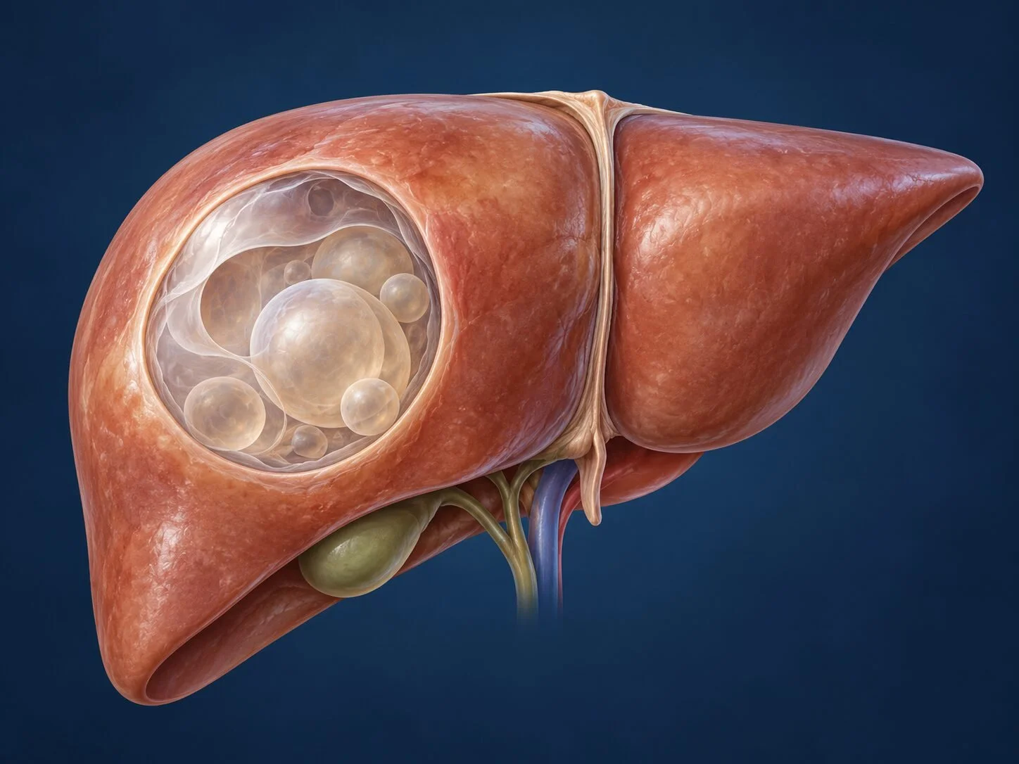
Hydatid disease is a parasitic infection of the liver caused by the larval stage of the tapeworm Echinococcus granulosus. It remains relevant in Cyprus, Greece and the wider Mediterranean, and it is the one cystic liver lesion where the distinction from a simple cyst genuinely changes management.
How it forms
The parasite lives its adult life in the intestine of dogs and other canines. Eggs are passed in dog faeces and contaminate pasture, vegetables, water and the animal's own coat. Sheep, goats and cattle ingest the eggs while grazing; cysts develop in their organs; and the cycle completes when a dog eats infected offal. Humans become involved by accident, swallowing eggs from contaminated hands, food or water, or through close contact with an infected dog.
Once swallowed, the egg hatches in the small intestine and the embryo crosses the bowel wall into the portal venous blood. The liver is the first filter it meets, which is why roughly two-thirds of human hydatid cysts occur there. Those that pass through lodge most often in the lung, and occasionally elsewhere.
In the liver the embryo develops slowly into a fluid-filled cyst with a characteristic structure: an inner germinal layer that produces the infective elements, a surrounding laminated membrane, and an outer fibrous capsule formed by the body's own reaction. The germinal layer buds off daughter cysts and protoscolices, which settle as "hydatid sand". Growth is slow, often a centimetre or less per year, so infection acquired in childhood may not be discovered until middle age.
Understanding this structure explains the central surgical principle: the cyst contents are both infective and capable of provoking a severe allergic reaction. Spillage during treatment can seed the abdomen with new cysts or trigger anaphylaxis. Every aspect of management is designed around not spilling.
How it is found
Many cysts are discovered incidentally on imaging. Others present when the cyst becomes large enough to cause symptoms, or when a complication occurs. A history of rural residence, farming, or contact with sheep-rearing and dogs supports the diagnosis but its absence does not exclude it.
Symptoms
often none for many years
dull right upper abdominal pain or a feeling of heaviness
a palpable mass when large
nausea, or fullness after small meals
Complications produce more dramatic presentations: rupture into the biliary tree causes jaundice, biliary colic and cholangitis; rupture into the abdominal cavity causes sudden pain and may provoke an allergic or anaphylactic reaction; secondary bacterial infection produces fever and the picture of an abscess; and a large cyst may compress bile ducts or blood vessels.
How the diagnosis is made
Ultrasound is the primary tool and is often diagnostic. Characteristic features include daughter cysts within the parent cyst, a detached and undulating inner membrane, and calcification of the wall. Ultrasound appearances are classified into recognised stages, which matters because the stage indicates whether the cyst is active, transitional or inactive.
CT and MRI add information about the number and position of cysts, their relationship to the vessels and bile ducts, and the presence of biliary communication — a crucial point before any intervention. MRCP is used when connection to the biliary tree is suspected.
Serological tests for Echinococcus support the diagnosis. They are not perfectly sensitive: a negative result does not exclude hydatid disease, particularly for calcified or inactive cysts, and a positive result can persist for years after successful treatment.
Diagnostic needle aspiration of a suspected hydatid cyst is avoided as a routine step, precisely because of the risk of spillage.
When treatment is needed
Not every hydatid cyst requires an operation. Small, entirely calcified, inactive cysts in a patient without symptoms may reasonably be watched, with imaging follow-up. Treatment is indicated for active or transitional cysts, large cysts, cysts causing symptoms, cysts that have communicated with the biliary tree, and those that are infected or at risk of rupture.
Treatment
Antiparasitic medication
Albendazole is given before and after intervention to reduce the viability of the parasite and lower the risk of recurrence if spillage occurs. It is occasionally used alone for small cysts or for patients unfit for surgery, but medication by itself cures a minority.
PAIR and percutaneous techniques
Puncture, Aspiration, Injection of a scolicidal agent, and Re-aspiration, performed under imaging guidance with antiparasitic cover. Suitable for selected cysts without daughter cysts and without biliary communication. Not appropriate where the cyst communicates with the bile ducts, because scolicidal agents can damage the biliary epithelium.
Surgery
Operative treatment ranges from conservative to radical. Conservative procedures remove the cyst contents and the germinal and laminated layers while leaving the outer fibrous capsule, which is then managed by deroofing, omentoplasty or drainage. Radical procedures remove the cyst with its capsule intact — pericystectomy — or, occasionally, require formal liver resection. Radical approaches carry a lower recurrence rate; the choice depends on the cyst's position, its relationship to major vessels and ducts, and the patient's fitness.
Throughout the operation the field is packed with scolicidal-soaked material and the cyst is isolated before opening, to contain any spillage. Where the cyst communicates with the biliary tree, the connection is identified and closed.
Selected cases are suitable for a laparoscopic approach. The decision rests on cyst position and the ability to maintain the same containment principles through small incisions rather than on the wish to avoid an open operation.
Recovery
Recovery depends on the procedure. After conservative laparoscopic surgery the stay is typically two to four days; after open pericystectomy or liver resection, longer. Albendazole is usually continued for a period after surgery. Bile leak is the most common specific complication and is managed according to its volume and duration.
Follow-up
Follow-up imaging is arranged over several years, because recurrence can appear late. Serology is used alongside imaging, bearing in mind that titres fall slowly after successful treatment.
When to seek a surgical opinion
Any cystic liver lesion with daughter cysts, a visible membrane, or wall calcification should be assessed by a surgeon familiar with hydatid disease before anything is done to it — and specifically before anyone puts a needle into it. In a region where the disease is endemic, that caution is worth exercising even when the appearances look straightforward.
Common questions
How did I catch this?
By swallowing microscopic eggs of a tapeworm, usually from food, water or soil contaminated by dog faeces, or from close contact with a dog whose coat carries them. The dogs themselves appear healthy. It is commonest in sheep-farming areas, where dogs are fed sheep offal, and infection frequently occurred in childhood, many years before the cyst was found.
Can I pass it to my family?
No. The parasite is not transmitted from person to person. Your relatives are at risk only if they have had the same environmental exposure, and screening them with an ultrasound is reasonable where that is the case.
Why can't the cyst simply be drained?
Because the fluid contains large numbers of infective particles, and spilling it inside the abdomen can seed new cysts throughout the cavity or, occasionally, cause a severe allergic reaction. Where drainage is appropriate, it is done under strict conditions with a parasite-killing agent injected first, and with medication given beforehand.
Do I have to have an operation?
Not always. Small, inactive, calcified cysts often need nothing but monitoring. Others are treated with medication alone, or by a needle technique under imaging guidance. Surgery is used for large, complicated or awkwardly placed cysts, and for those that have leaked into the bile ducts.
How long do I take the tablets for?
Typically for a period before any procedure and for some months afterwards, to reduce the risk of spillage causing new cysts. The course is longer than most people expect, and liver function and blood counts are monitored while you are on it.
Can it come back?
Yes, which is why follow-up with scans and blood tests continues for several years. Recurrence is more likely after incomplete removal or spillage, and less likely after a complete removal of the cyst and its lining.
Is it dangerous?
It is usually slow-growing and many people live with one for years without knowing. The complications that matter are rupture into the bile ducts, causing jaundice and infection, rupture into the abdomen or chest, and infection of the cyst. These are the reasons treatment is recommended for active cysts rather than simply watching them.
Related conditions
Other conditions of the liver covered on this site:
This page provides general information and does not replace an individual medical consultation. Assessment and treatment are decided for each patient after review of their history, examination and imaging.
