

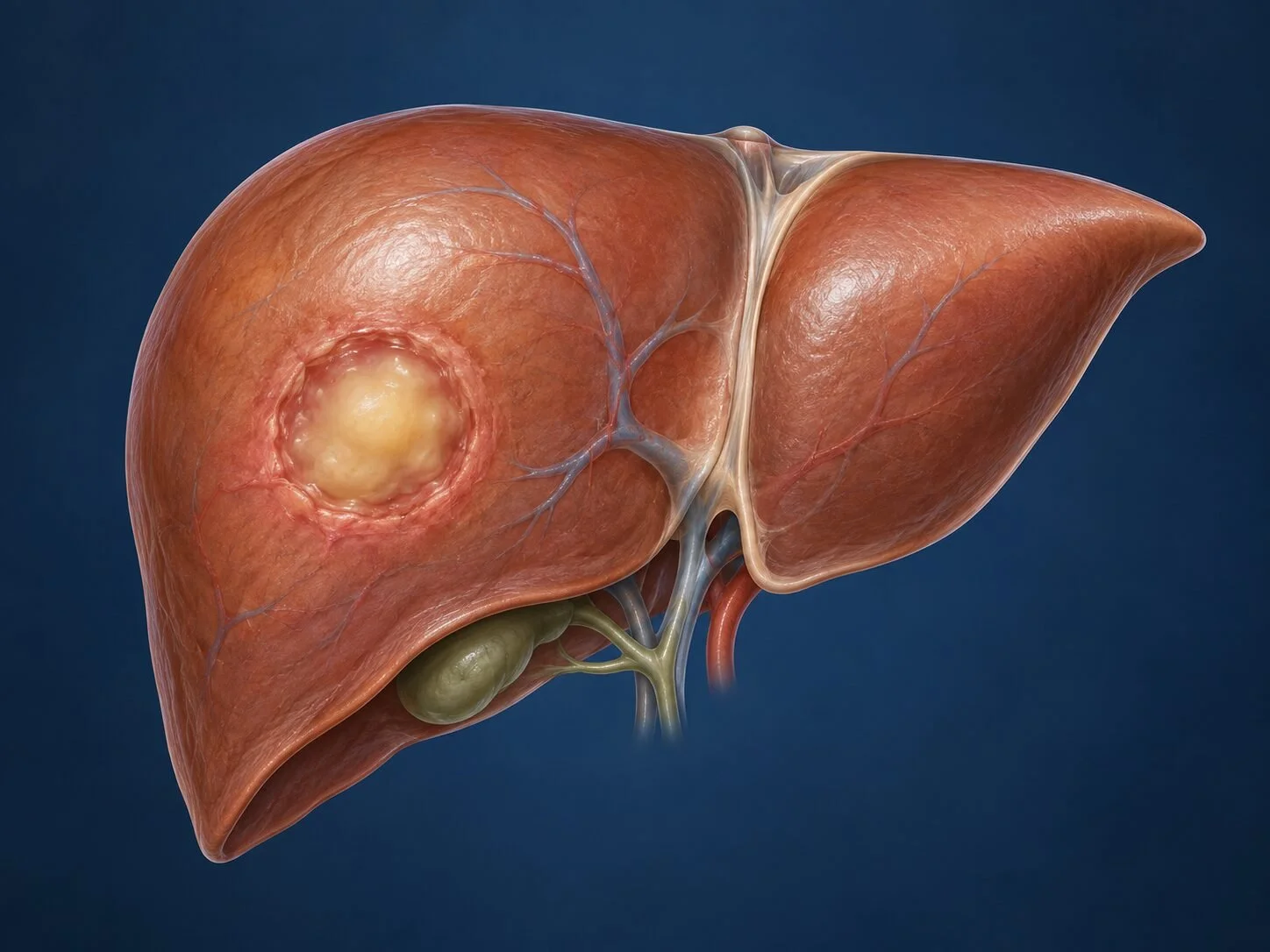
A liver abscess is a collection of pus within the liver. Unlike most of the other lesions described in this section, it is an acute illness rather than an incidental finding, and it requires prompt treatment. With modern imaging, drainage techniques and antibiotics, the outlook is good — but delay is what causes harm.
How it forms
The liver receives blood from two sources and bile from the biliary tree, and an abscess can arise through any of these routes. The route determines the likely organism and, importantly, points to an underlying cause that also needs treating.
Biliary — the commonest route. Obstruction of the bile ducts by a stone, a stricture or a tumour causes stasis and ascending infection (cholangitis), which seeds the liver. Abscesses arising this way are often multiple.
Portal venous — infection from an intra-abdominal source such as appendicitis, diverticulitis or an inflamed bowel travels in the portal vein to the liver.
Arterial — bloodstream infection from a distant site seeds the liver via the hepatic artery.
Direct spread from an adjacent structure, or following trauma or an intervention on the liver.
Whatever the route, bacteria establish themselves in liver tissue, provoke an inflammatory response, and the tissue liquefies into a cavity of pus walled off by the body's reaction.
A separate entity is the amoebic liver abscess, caused by Entamoeba histolytica reaching the liver from the bowel. It is uncommon in Cyprus but relevant in patients who have travelled to or come from endemic regions, and it is treated primarily with medication rather than drainage.
The single most important consequence of this mechanism: a liver abscess is almost always a symptom of something else. Finding the abscess is only half the diagnosis.
Symptoms
fever, often with rigors and night sweats
pain in the right upper abdomen, sometimes referred to the right shoulder
malaise, loss of appetite, weight loss
nausea and vomiting
jaundice, particularly when the cause is biliary
The presentation can be surprisingly indolent, especially in older patients, where unexplained fever and weight loss over several weeks may be the only features.
How the diagnosis is made
Blood tests show raised inflammatory markers and white cell count, often with abnormal liver function tests. Blood cultures are taken before antibiotics are started whenever possible, because identifying the organism guides treatment.
Ultrasound usually identifies the collection. Contrast-enhanced CT defines it more precisely — its size, number, position and relationship to the bile ducts and vessels — and, critically, looks for the underlying cause: gallstones, biliary obstruction, diverticulitis, appendicitis or a colonic tumour.
Aspiration of the collection provides material for culture and is often therapeutic at the same time. Serology is requested where amoebic abscess is a possibility.
A point worth emphasising: in a patient over fifty with a liver abscess of portal origin and no obvious source, the colon should be examined. An underlying colorectal cancer is an uncommon but well-recognised cause, and missing it is the serious error.
Treatment
Antibiotics
Broad-spectrum intravenous antibiotics are started promptly and then narrowed once cultures identify the organism. Treatment continues for several weeks, often transitioning to oral therapy, with the duration guided by clinical response and imaging.
Drainage
Most abscesses of any size require drainage in addition to antibiotics. Percutaneous drainage under ultrasound or CT guidance is the standard approach — either aspiration alone for smaller collections, or placement of a drainage catheter for larger ones. This is effective in the great majority of cases.
Surgery
Surgical drainage is reserved for abscesses that cannot be reached percutaneously, those that fail to resolve, multiloculated collections, rupture into the abdominal cavity, or cases where an operation is needed anyway to treat the underlying cause. The approach may be laparoscopic or open.
Treating the cause
This is not an optional extra. Biliary obstruction is relieved, usually endoscopically; gallstones are dealt with; an underlying bowel condition is treated. An abscess drained without addressing its source tends to return.
Recovery
Recovery is gradual. Fever typically settles within days of adequate drainage. The drain remains until output falls and imaging confirms resolution, which may take one to three weeks. Fatigue often persists for several weeks beyond that, and patients should expect a recovery measured in weeks rather than days. Repeat imaging confirms the cavity has closed.
Follow-up
Follow-up covers two things: confirming the abscess has resolved, and completing the investigation and treatment of the underlying cause. Where a colonic source was suspected, colonoscopy is arranged once the acute illness has settled.
When to seek medical attention
Fever with right upper abdominal pain is not something to wait out, particularly in anyone with known gallstones, recent abdominal infection or recent biliary intervention. This is an acute condition, and the sensible course is prompt assessment rather than a routine appointment.
Common questions
How does an abscess form in the liver?
Almost always because bacteria reach the liver from somewhere else. The commonest route is from an infected bile duct, usually obstructed by a stone or a stricture. Another is through the portal vein, which drains the intestine, so appendicitis, diverticulitis or an inflamed bowel can seed the liver. Occasionally it comes through the bloodstream from an infection elsewhere entirely.
Why do I need scans looking at the rest of my abdomen?
Because a liver abscess is usually a consequence of something else, and finding the source is as important as treating the abscess. Bile duct stones, diverticulitis, appendicitis and bowel disease all need identifying and treating, or the abscess returns.
Why is a colonoscopy sometimes recommended afterwards?
Because an underlying bowel cancer can occasionally be the source, particularly where a specific organism is grown or where no other cause is found. It is a precaution rather than an expectation, and it is worth completing.
Can antibiotics alone cure it?
Small abscesses often resolve with antibiotics alone. Larger ones generally need draining, because pus within a wall has a poor blood supply, so antibiotics reach it only in low concentrations and cannot clear it. Draining it through the skin under scan guidance avoids an operation in the great majority.
How long will I need antibiotics?
Longer than for most infections — commonly several weeks, often beginning intravenously and continuing by mouth, with the duration guided by your symptoms, blood tests and repeat scans. Stopping early is a recognised cause of recurrence.
Will I need an operation?
Usually not. Most are drained with a needle and tube placed through the skin. Surgery is reserved for abscesses that cannot be reached safely, those that are multiple or thick-walled, those that have ruptured, and cases where the underlying cause needs operating on anyway.
I have recently travelled abroad. Does that change anything?
It may. An amoebic abscess, caused by a parasite rather than bacteria, is treated with a specific drug rather than by drainage, and it responds rapidly. Travel to areas where this is common, and a blood test for the parasite, both form part of the assessment, so mention any travel history.
How long will recovery take?
Longer than people expect. Fever usually settles within days of adequate drainage, but tiredness commonly persists for several weeks, and the drain may stay in for one to three weeks. A follow-up scan confirms the cavity has closed before treatment is stopped.
Related conditions
Other conditions of the liver covered on this site:
This page provides general information and does not replace an individual medical consultation. Assessment and treatment are decided for each patient after review of their history, examination and imaging.
