

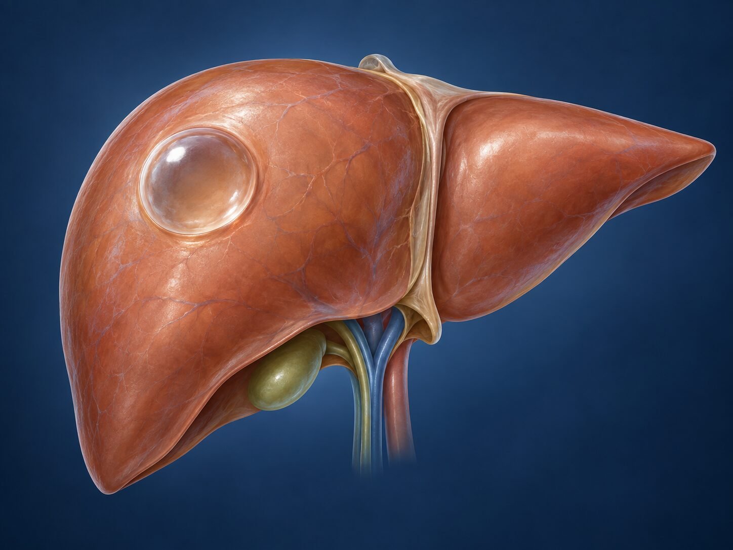
Simple liver cysts are fluid-filled spaces within the liver tissue. They are among the most common findings on abdominal imaging, are almost always benign, and in the great majority of people never cause a problem or require treatment.
How they form
Simple liver cysts are developmental rather than acquired. During formation of the biliary system before birth, small clusters of bile duct cells fail to connect with the rest of the biliary tree and become isolated within the liver tissue. These isolated groups retain the lining cells of a bile duct, and those cells continue to do what they are designed to do — secrete fluid. With nowhere for the fluid to drain, it accumulates slowly, over decades, and the space gradually enlarges into a cyst.
This explains several things patients often ask about. It explains why cysts are lined by biliary-type epithelium when examined under the microscope; why they contain clear, watery fluid rather than blood or pus; why they are found more often with increasing age, despite having been present since birth; and why they grow so slowly that most never reach a size that causes trouble.
It also explains what does not cause them. Simple liver cysts are not caused by diet, alcohol, medication, infection or anything you have done. They are not a form of liver damage and they do not turn into cancer. Hormonal influence may play some part in growth, which is one explanation offered for why larger symptomatic cysts are seen more often in women, but the cyst itself was not caused by that.
Polycystic liver disease is a different matter. There the cause is genetic — either an inherited condition affecting the liver alone, or the liver manifestation of autosomal dominant polycystic kidney disease. Numerous cysts form throughout the liver by the same mechanism, but far more of them, and the condition runs in families.
How they are usually found
Most simple cysts are discovered incidentally — on an ultrasound, CT or MRI performed for an unrelated reason. They become more common with age and are seen more often in women. A single cyst is typical; some people have several.
A separate condition, polycystic liver disease, causes numerous cysts throughout the liver and is usually inherited. It is assessed and managed differently from an isolated simple cyst.
Symptoms
Small cysts cause no symptoms at all. When a cyst becomes large, it can produce:
a dull ache or feeling of fullness in the upper right abdomen
early satiety — feeling full soon after starting to eat
discomfort on bending or lying on one side
rarely, a palpable swelling
Sudden severe pain is uncommon and may indicate bleeding into a cyst, rupture or infection. These situations need prompt assessment.
How the diagnosis is made
Ultrasound is usually sufficient to identify a simple cyst: it appears as a thin-walled structure with clear fluid and no solid component. When the appearance is not entirely typical, contrast-enhanced CT or MRI is used to characterise it more precisely.
The purpose of imaging is not only to confirm the cyst but to exclude the small number of cystic lesions that are not simple cysts — hydatid disease, a cystic tumour such as a mucinous cystic neoplasm, or an abscess. Features that prompt closer scrutiny include a thick or irregular wall, internal septations, solid nodules, calcification, or enhancement after contrast.
Blood tests are normal in simple cysts. Serology for Echinococcus is requested when hydatid disease is a possibility, which is relevant in Cyprus and the wider Mediterranean.
When treatment is needed — and when it is not
The default for a typical simple cyst causing no symptoms is no treatment. Repeated scanning is generally unnecessary once the diagnosis is secure, although a single follow-up scan is sometimes arranged to confirm stability.
Treatment is considered when:
symptoms are genuinely attributable to the cyst rather than to another cause
the cyst is enlarging significantly over time
imaging leaves doubt about whether the lesion is truly a simple cyst
a complication has occurred — haemorrhage, infection or rupture
It is worth being clear about the second point: upper abdominal symptoms are common and have many causes. Before attributing them to a cyst, other explanations are considered, because removing a cyst will not relieve pain that was never coming from it.
Treatment options
Laparoscopic fenestration (deroofing)
The established surgical treatment for a symptomatic simple cyst. Through several small incisions, the exposed dome of the cyst is excised, allowing the fluid to drain freely into the abdominal cavity, where it is absorbed. The cyst lining that remains against the liver is inspected and biopsied when appropriate.
Recurrence rates are low when an adequate window is created. The operation is usually completed laparoscopically; conversion to an open approach is occasionally necessary depending on the position of the cyst, particularly for lesions in the posterior segments.
Aspiration and sclerotherapy
Drainage with a needle under imaging guidance, sometimes followed by injection of a sclerosing agent. Simple aspiration alone is followed by refilling in most cases and is therefore used mainly as a diagnostic step or as temporary relief. Aspiration is avoided when hydatid disease has not been excluded.
Liver resection
Reserved for situations where a cystic tumour cannot be excluded, or where a cyst is unsuitable for fenestration. The extent of resection depends on the position of the lesion.
Recovery
After laparoscopic fenestration most people stay in hospital one or two nights. Discomfort at the port sites settles over several days, and shoulder-tip discomfort from the gas used during laparoscopy is common for a day or two. Ordinary activity resumes within one to two weeks and full activity, including lifting, by about four weeks. Symptom relief, when the cyst was genuinely the cause, is usually immediate.
Follow-up
Routine long-term surveillance is not required for a treated simple cyst. Where a cystic neoplasm was suspected or confirmed, follow-up is guided by the final histology and arranged accordingly.
When to seek a surgical opinion
A surgical opinion is reasonable when a cyst is causing symptoms that interfere with daily life, when a cyst is growing, or when imaging has raised any question about the nature of the lesion. An incidental small cyst with classic features on a good-quality scan does not usually need a surgeon at all, and saying so is part of the assessment.
Common questions
I have a cyst on my liver. Is it cancer?
Almost certainly not. A simple liver cyst is a fluid-filled sac with a thin wall and clear contents, and it has no potential to become cancer. On a good-quality ultrasound or CT the appearance is characteristic, and once those features are confirmed no further concern is warranted.
Does it need to be removed?
Only if it is causing symptoms. Most liver cysts are found by accident and cause nothing at all, and the right treatment for those is none. Removing a cyst that is not troubling you exposes you to the risks of an operation for no benefit.
Will it keep growing?
Some enlarge slowly over years and many stay the same. Growth alone is not a reason for treatment; what matters is whether it produces symptoms. A cyst that changes in character — developing thick walls, solid areas or internal divisions — is a different matter and needs reassessment.
Why not just drain it with a needle?
Because the lining continues to produce fluid, so a cyst that is simply aspirated refills within weeks in the great majority of cases. Draining and then instilling an agent to destroy the lining works better, and surgically removing the roof of the cyst so it drains permanently into the abdomen works best of all.
Could it burst?
This is very rare. Simple cysts do not usually rupture, and the thin wall means that even a large one is not under significant pressure. Sudden severe pain in a known cyst should be assessed, as bleeding into a cyst can occur, but this is uncommon.
I have been told I have polycystic liver disease. Is that the same thing?
No. That is an inherited condition with large numbers of cysts throughout the liver, often alongside kidney cysts, and it is managed differently — the aim is to reduce the overall volume rather than deal with one cyst. It also warrants assessment of your kidneys and consideration of testing for relatives.
Do I need repeat scans?
Usually not, once a cyst has been confidently identified as simple. A single repeat scan after an interval is sometimes arranged for reassurance, after which no further imaging is needed unless symptoms develop.
Related conditions
Other conditions of the liver covered on this site:
This page provides general information and does not replace an individual medical consultation. Assessment and treatment are decided for each patient after review of their history, examination and imaging.
