

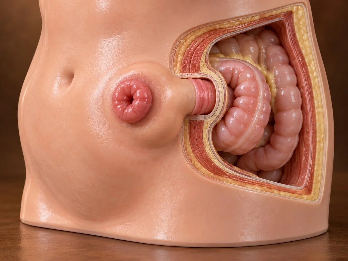
A parastomal hernia is a hernia through the abdominal wall defect created for a stoma. It is extremely common — so common that it has been described, not unfairly, as an inevitable consequence of forming a stoma rather than a complication of it. Over half of patients develop one within two years, and the proportion continues to rise thereafter. For many it causes no more than an awkward bulge; for others it makes the appliance leak, which is a considerable daily burden. Repair is difficult and recurrence is high, which is why prevention has become the principal focus.
How they form
The problem is intrinsic to what a stoma is. Forming one requires a permanent hole in the abdominal wall through which bowel passes, and that hole is a hernia defect by definition. Several forces then act to enlarge it:
The aperture cannot be closed. It must remain large enough to avoid obstructing the bowel and its blood supply, so a tension-free seal is impossible.
Continuous pressure. Intra-abdominal pressure acts on the aperture with every cough, strain and movement, gradually stretching it.
The bowel itself moves with peristalsis, working against the rim.
Impaired healing at the interface — the junction between bowel serosa and fascia never forms a strong union.
Divided muscle and nerves. Bringing the stoma through the rectus muscle helps, but any aperture interrupts the structural continuity of the wall.
Risk is increased by obesity, which is much the most important factor, by siting the stoma lateral to the rectus muscle rather than through it, by an aperture made larger than necessary, by emergency formation, by wound infection, by chronic cough or constipation, by steroids and malnutrition, and by the underlying connective tissue weakness common to all hernia patients. Colostomies carry a higher rate than ileostomies, and end stomas a higher rate than loop stomas.
Hernias are classified by the European Hernia Society by size and by whether a concurrent incisional hernia is present, which matters because the two frequently coexist and should be repaired together.
Symptoms
a bulge around the stoma, increasing on standing or straining
difficulty getting the appliance to stick, and leakage — usually the symptom that matters most to the patient, because it affects every day and every social situation
skin excoriation from leakage
discomfort, dragging, and a sense of the stoma sitting on a mound
difficulty with irrigation in colostomy patients
problems with clothing and body image
intermittent colicky pain and poor stoma output, from partial obstruction
Emergency features: a tense, painful bulge with no stoma output, vomiting and distension, indicating obstruction; a dusky or black stoma, indicating ischaemia. Both require immediate assessment. Strangulation is less common than in other hernias but does occur.
How the diagnosis is made
Examination with the patient standing and straining, and a careful history about appliance leakage, which is the symptom that determines whether repair is worthwhile.
CT is the investigation of choice and should be performed with the patient straining. It confirms the hernia, measures the defect, identifies the contents, and — importantly — identifies any coexisting incisional hernia in the midline, which is present in a large proportion of these patients and must be included in the surgical plan.
Treatment
When not to operate
This is a genuine and defensible position. Many parastomal hernias cause an unsightly bulge but no functional problem, and repair carries a substantial recurrence rate and real complication risk. Conservative management — a well-fitted appliance, a support garment or hernia belt with an aperture, specialist stoma nurse input, weight loss, and treatment of cough and constipation — is appropriate for many patients and is not a second-best option.
Specialist stoma nursing is the single most valuable intervention for a leaking appliance and is frequently under-used before surgery is contemplated.
When to repair
obstruction or strangulation — emergency
persistent leakage not solved by appliance adjustment and stoma nursing
intractable pain
a very large hernia limiting daily life
skin ulceration
at the time of another abdominal operation, when the hernia can be addressed concurrently
The operations
Suture repair alone — narrowing the aperture with stitches. Simple, but recurrence rates are very high, in the region of half to two thirds. It is now reserved for patients unfit for anything more.
Mesh repair — the standard. Several configurations exist:
Sugarbaker technique — the bowel is lateralised and a mesh placed over both the defect and the bowel, which passes between mesh and abdominal wall before turning. This creates a valve-like angulation and has among the lowest recurrence rates.
Keyhole technique — a mesh with a central hole through which the bowel passes. Simpler, but the hole tends to stretch and recurrence is higher.
Sandwich and modified techniques, combining elements of both.
Retromuscular (Pauli) repair — a retromuscular Sugarbaker configuration, increasingly favoured, placing mesh outside the peritoneal cavity and away from bowel.
Stoma relocation — moving the stoma to the other side, with repair of the original site. It simply creates a new aperture at a new site, which carries its own risk of hernia, and is generally reserved for situations where the current site is unusable.
Stoma reversal — where the stoma is potentially reversible, this is the definitive solution and should be considered before embarking on repair.
Laparoscopic and robotic approaches are widely used and reduce wound complications, which matters in a field adjacent to a stoma.
Prevention
Because repair is difficult, prevention has received considerable attention, and two measures are supported:
Prophylactic mesh at the time of stoma formation — placing a mesh in the retromuscular or preperitoneal plane around the stoma when it is created. Multiple trials and meta-analyses show a reduction in parastomal hernia, and although results have been mixed and the effect size debated, it is recommended in guidelines for permanent end colostomy.
Careful stoma siting — marked preoperatively by a stoma nurse with the patient sitting, standing and lying, brought through the rectus muscle, away from scars, skin creases, the belt line and bony prominences, and visible to the patient. This is a genuinely important step and is sometimes omitted in emergency surgery, with lasting consequences.
Recovery
Recovery depends on the approach. Laparoscopic repair typically involves three to five days in hospital; open repair with reconstruction longer. A support garment is usually advised for some months. Return to full activity is six to eight weeks for open repair.
Seroma around the repair is common and settles. Recurrence is the principal problem, and patients should be given a realistic figure rather than an optimistic one — even with modern mesh techniques it remains meaningfully higher than for other hernias.
When to seek an opinion
Persistent appliance leakage, a bulge that is enlarging, or discomfort around the stoma. See a stoma nurse first; many problems are solved at that level. No output from the stoma with pain, vomiting or distension, or a stoma that looks dusky or black, requires emergency assessment.
Common questions
Is this my fault?
No. A hernia around a stoma is extremely common — more than half of people develop one within two years — because forming a stoma necessarily means leaving a permanent opening in the abdominal wall that cannot be closed. It is better thought of as an expected consequence than as something that went wrong.
Do I need an operation?
Not necessarily, and many people do better without one. If the bulge is mainly cosmetic and the appliance sticks well, a support garment, weight control and good stoma nursing may be all that is needed. Surgery is worth considering when the appliance leaks despite expert adjustment, when there is significant pain, or when the hernia is very large.
My bag keeps leaking. What can be done first?
A specialist stoma nurse is the most valuable person to see, and often solves the problem without surgery — different appliance shapes, convex flanges, seals, belts and support garments all make a considerable difference on an uneven surface. This should always be tried thoroughly before an operation is considered.
How likely is it to come back after repair?
More likely than for other hernias, and you should be given a frank answer. Repairs using stitches alone fail in the majority. Modern mesh repairs are substantially better but recurrence remains a real possibility. This is why the decision to operate is weighed carefully against how much trouble the hernia is actually causing.
Could the stoma be moved to the other side?
It can, but moving it simply creates a new opening elsewhere, which carries its own risk of a hernia forming there, and leaves the old site to be repaired as well. It is generally reserved for situations where the current site cannot be made to work at all.
Could my stoma be reversed instead?
If your stoma is potentially reversible, this is worth asking about, because it solves the problem definitively rather than patching it. Whether it is possible depends on the original operation, the state of the remaining bowel and your general fitness.
Could this have been prevented?
Partly. Placing a mesh around the stoma when it is first formed reduces the rate, and is now recommended for permanent colostomies. Careful marking of the site beforehand by a stoma nurse, with you sitting, standing and lying down, also matters — though in emergency surgery there is often no opportunity for either.
Is it dangerous?
Usually not. Serious trapping of bowel is less common than with other hernias because the opening is relatively wide. It should nonetheless be taken seriously if the stoma stops working, you develop pain and vomiting, or the stoma changes colour — those need immediate attention.
Related conditions
Other conditions of the hernias & abdominal wall covered on this site:
This page provides general information and does not replace an individual medical consultation. Assessment and treatment are decided for each patient after review of their history, examination and imaging.
