

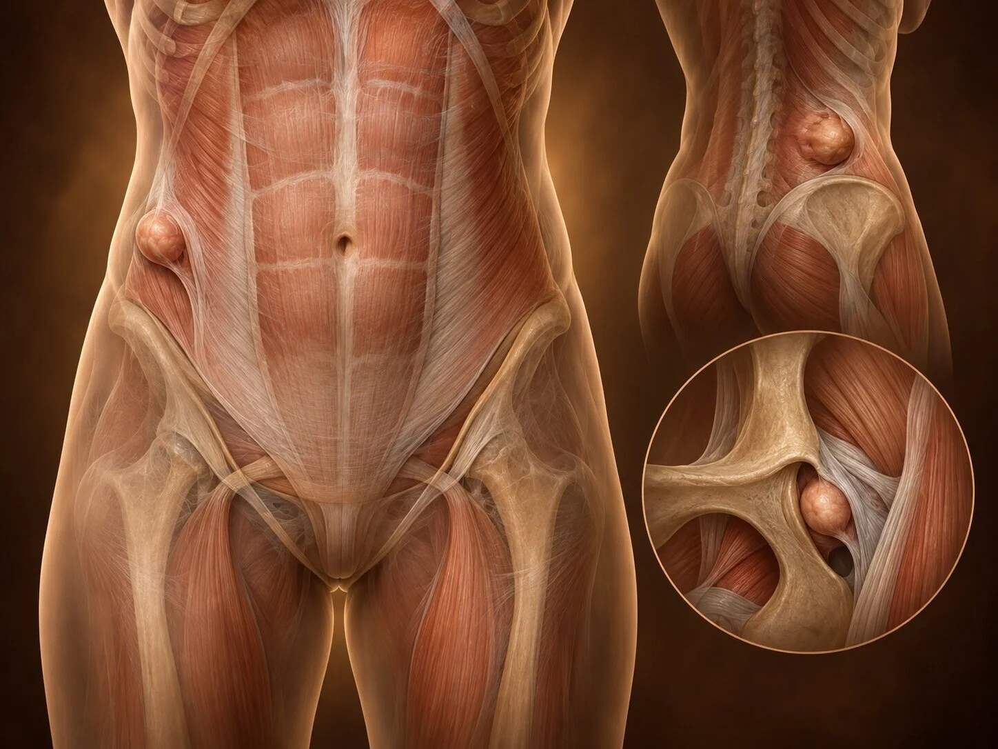
Beyond the familiar groin, umbilical and incisional hernias lies a group of uncommon hernias that share one important characteristic: they are difficult to see and feel, so they are frequently missed. Several present for the first time as bowel obstruction, sometimes after months of being investigated for something else. They are worth knowing about precisely because the diagnosis depends on someone thinking of them, and because a CT scan will show them when examination will not.
Spigelian hernia
How it forms
The Spigelian hernia occurs through the semilunar line — the curved junction where the muscle fibres of transversus abdominis become aponeurosis, at the lateral border of the rectus sheath. The vulnerable point is where this line crosses the arcuate line, below which the posterior rectus sheath is absent and the abdominal wall is correspondingly thinner. This area, a few centimetres above and lateral to the pubic tubercle, is the "Spigelian belt", and almost all such hernias occur there.
What makes it deceptive is that the defect lies in the transversus and internal oblique layers, while the external oblique aponeurosis remains intact over it. The hernia therefore spreads out beneath that intact layer — it is interparietal, lying within the abdominal wall rather than pushing out through the skin. There may be no visible lump at all.
Because the defect is small with a firm fibrous rim, the risk of incarceration and strangulation is high — considerably higher than for an ordinary inguinal hernia.
Presentation and diagnosis
Intermittent, poorly localised pain in the lower lateral abdomen, often for months, usually worse on straining and relieved by lying down. A lump is palpable in only a minority, and then best felt with the patient straining. Many are diagnosed only when they obstruct.
Ultrasound with straining, and CT, are diagnostic. CT is the more reliable and also identifies the contents.
Treatment
All Spigelian hernias should be repaired because of the high risk of strangulation. Repair is with mesh, open or — more commonly now — laparoscopically or robotically, which has the advantage of identifying the defect accurately from within and placing mesh with wide overlap.
Lumbar hernia
How it forms
Two anatomical triangles in the posterior abdominal wall are natural weak points:
Superior (Grynfeltt-Lesshaft) triangle — bounded by the twelfth rib, internal oblique and sacrospinalis. The larger of the two and the commoner site.
Inferior (Petit) triangle — bounded by the iliac crest, latissimus dorsi and external oblique.
Lumbar hernias are congenital in a small proportion, or acquired — following flank incisions (nephrectomy, aortic surgery, iliac crest bone graft harvest), trauma, or infection. Incisional lumbar hernias after flank surgery are the commonest type in practice, and they are frequently large and diffuse, complicated by denervation of the flank muscles from the original incision, which leaves a bulging, weakened area that is partly hernia and partly muscle paralysis.
Presentation and diagnosis
A soft flank bulge, back or flank pain, and a sensation of dragging. The bulge may be obvious on standing and disappear on lying. Strangulation is uncommon because the defects are typically broad, but obstruction does occur.
The essential diagnostic question, which CT answers, is whether the bulge is a true hernia with a fascial defect or a denervation bulge with an intact wall. The two look similar and are treated entirely differently — operating on a denervation bulge achieves little.
Treatment
Repair with mesh, with wide overlap fixed to the bony landmarks — rib, iliac crest, and the muscles — since there is limited fascia to anchor to on some sides. Laparoscopic and robotic retroperitoneal approaches are well suited to this. Results are less predictable than for anterior hernias, and recurrence is higher, particularly where denervation coexists.
Obturator hernia
How it forms
The obturator canal is a short tunnel in the pelvic bone transmitting the obturator nerve and vessels. It is normally plugged by fat and connective tissue. With marked weight loss, that fat is lost and the canal opens. The hernia then passes through a rigid, bony, unyielding ring.
It has a characteristic patient: the elderly, thin, multiparous woman — "the little old lady's hernia" — often with chronic cough or constipation. A wider female pelvis and pregnancy-related laxity explain the sex distribution.
Because the ring is bony and rigid, strangulation is very common, and the mortality is the highest of any hernia — both because of the delay in diagnosis and because of the frailty of the patients.
Presentation and diagnosis
There is almost never a palpable lump. The presentation is small bowel obstruction in a thin elderly woman, often with a history of previous self-resolving episodes.
The one distinctive clinical sign is the Howship-Romberg sign: pain along the inner thigh and medial knee, caused by compression of the obturator nerve, characteristically worsened by extension, abduction or internal rotation of the hip and relieved by flexion. It is present in a significant minority and is frequently attributed to arthritis. The Hannington-Kiff sign — absent adductor reflex with a preserved knee jerk — is more specific but less often tested.
CT is diagnostic and has transformed the outlook for this condition, which historically was diagnosed at laparotomy. Any elderly thin woman with unexplained small bowel obstruction should have her obturator canals examined on the scan.
Richter's hernias are common here, so bowel function may be preserved while the trapped bowel wall dies.
Treatment
Urgent surgery. The approach may be open (transabdominal, preperitoneal or via the thigh) or laparoscopic. The defect is closed with mesh where the field is clean. Bowel resection is frequently required because of delayed presentation.
Other rare hernias
Sciatic hernia — through the greater or lesser sciatic foramen. Extremely rare; presents with buttock pain, sciatic symptoms or obstruction.
Perineal hernia — through the pelvic floor, usually after abdominoperineal excision of the rectum or pelvic exenteration. Increasingly seen as more radical pelvic surgery is performed, and repaired with mesh or flap reconstruction.
Littré's hernia — containing a Meckel's diverticulum.
Amyand's hernia — containing the appendix, which may be normal or inflamed; management depends on which.
Internal hernias — through defects within the abdomen rather than the wall: paraduodenal, foramen of Winslow, transmesenteric, and — importantly — through mesenteric defects after Roux-en-Y gastric bypass. These present with intermittent or acute obstruction, may have normal-looking imaging between episodes, and are a recognised cause of serious delayed morbidity after bariatric surgery. Persistent abdominal pain after gastric bypass should be taken seriously and investigated, not attributed to diet.
Traumatic diaphragmatic hernia — abdominal contents entering the chest through a tear, which may present years after the original injury.
The common thread
These hernias share a diagnostic lesson worth stating explicitly: in unexplained intestinal obstruction, or in unexplained chronic abdominal pain with a normal examination, a CT scan looking specifically at all the hernial orifices is the investigation that makes the diagnosis. Almost every delayed diagnosis in this group reflects the condition simply not being considered.
When to seek an opinion
Persistent, unexplained abdominal or flank pain, particularly if intermittent and related to straining. Episodes of colicky pain with vomiting that settle on their own. Pain down the inner thigh in an elderly woman with abdominal symptoms. Vomiting with abdominal distension and inability to pass wind requires emergency assessment.
Common questions
How can I have a hernia with no lump?
Several of these hernias lie within the layers of the abdominal wall or deep in the pelvis, so nothing pushes out to the skin. A Spigelian hernia spreads out underneath an intact outer layer; an obturator hernia sits inside the bony pelvis. This is why they are missed on examination and why a CT scan is what makes the diagnosis.
I have had intermittent pain for months and everything has been normal. Could this be it?
It is worth considering, particularly if the pain is related to straining, comes in episodes, and settles when you lie down. These hernias characteristically cause months of intermittent symptoms before they are found, and a CT scan performed with this specifically in mind will show them.
Why is an obturator hernia so serious?
Because it passes through a rigid bony channel that cannot stretch, so trapped bowel loses its blood supply quickly; because there is no lump to see; and because it occurs in thin, elderly patients who tolerate a delayed emergency operation poorly. The combination is what gives it the worst outcomes of any hernia, and the answer is thinking of it early.
I have pain down my inner thigh. What has that to do with my abdomen?
A hernia passing through the obturator canal compresses the nerve running through it, and that nerve supplies the inner thigh and knee. Inner thigh pain that worsens when the hip is extended or turned inward, in someone with abdominal symptoms, is a recognised clue — and one that is often put down to arthritis.
My flank bulges after kidney surgery. Is that a hernia?
It may be, or it may be that the nerves supplying the muscles were divided during the original incision, leaving the muscle weak and bulging without any actual hole. A CT scan distinguishes the two, and this matters because an operation helps a true hernia but achieves little for a paralysed muscle.
Do rare hernias need repairing?
Spigelian and obturator hernias should be repaired because of the high risk of trapped bowel. Lumbar and perineal hernias are repaired for symptoms, and their repair is less predictable. In each case the decision is individual rather than automatic.
I have ongoing pain after gastric bypass surgery. Should I be concerned?
Yes, it is worth investigating properly. Bowel can herniate through gaps between the loops rearranged during the operation, and this can cause intermittent pain with normal scans between episodes. Persistent or recurrent abdominal pain after bypass surgery should be assessed by a surgeon familiar with the operation rather than attributed to diet.
Related conditions
Other conditions of the hernias & abdominal wall covered on this site:
This page provides general information and does not replace an individual medical consultation. Assessment and treatment are decided for each patient after review of their history, examination and imaging.
