

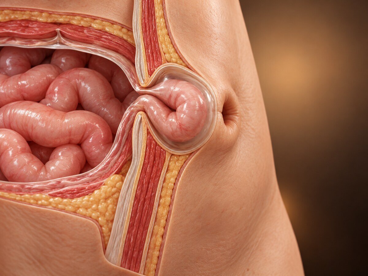
An umbilical hernia is a protrusion through the natural weakness at the navel. It is one of the most familiar of all hernias — obvious to the patient, easily examined, and often present for years before anything is done about it. Two things are worth knowing from the start: umbilical hernias in children behave entirely differently from those in adults, and in adults these hernias have a relatively narrow neck, which makes them more likely than a large incisional hernia to trap their contents.
How they form
The umbilicus is the site where the umbilical vessels passed through the abdominal wall in fetal life. After birth the ring contracts and the defect closes, leaving a scarred area covered only by skin, scar tissue and peritoneum, with no muscle. It is a natural weak point in an otherwise continuous wall.
In infants, failure of the ring to close completely leaves a true umbilical hernia. It is very common, particularly in premature and low-birthweight babies, and in certain ethnic groups. The great majority close spontaneously by the age of four or five, and surgery is not usually considered before then. Strangulation in childhood is rare.
In adults, hernias here are usually paraumbilical — occurring through the weakened tissue immediately above or below the umbilical ring rather than through the ring itself. They are acquired. They do not resolve spontaneously, and they enlarge.
Adult hernias at this site are driven by sustained raised intra-abdominal pressure acting on a connective tissue that is often inherently weak. The recognised contributors are obesity (much the most important), pregnancy, ascites from liver disease, chronic cough, constipation and straining, and previous surgery or laparoscopic port sites at the umbilicus.
As with other hernias, there is an underlying collagen abnormality in many patients — an altered balance of type I to type III collagen — which is why suture repair alone fails more often than expected and why mesh reinforcement improves durability.
Why they trap their contents
The defect typically has a firm fibrous rim and a relatively narrow neck compared with the volume of the sac. Omentum, and sometimes bowel, enters and swells, and cannot easily return. Incarceration is common; strangulation, though less common, occurs more readily than in a large incisional hernia. The contents are most often omentum, which when trapped causes localised pain and a tender lump without obstruction.
Umbilical hernia in liver disease
This warrants specific mention because it is managed differently and badly handled it can be lethal. In patients with cirrhosis and ascites, the fluid produces continuous pressure, the hernia enlarges, the overlying skin becomes stretched and thin, and the hernia may ulcerate and rupture, leaking ascites — a situation with a high mortality from infection. Elective repair in these patients, with the ascites and liver disease first controlled medically, is considerably safer than emergency repair of a ruptured hernia. Traditional advice to avoid operating on these patients has been revised.
Symptoms
a lump at or beside the navel, more prominent on standing, coughing or straining
aching or dragging discomfort, often worse at the end of the day
a lump that may or may not be reducible
skin changes over a large hernia — thinning, discolouration, or ulceration
in children, a soft bulge that becomes prominent when crying and reduces easily — usually painless
Emergency features: the lump becoming painful, firm and irreducible; skin redness over it; vomiting, abdominal distension and absolute constipation. Any of these requires immediate assessment.
How the diagnosis is made
Usually clinically. Ultrasound confirms the defect and its contents where examination is difficult. CT is used for large or complex hernias, to size the defect, assess the contents, plan the repair, and identify additional defects along the midline — which are common and easily missed on examination alone (a "Swiss cheese" pattern of multiple small defects).
Treatment
Children
Observation until the age of four or five, since most close by themselves. Repair is considered for hernias persisting beyond that age, very large defects, or the rare episode of incarceration. Taping or strapping is ineffective.
Adults
Adult hernias at this site do not resolve and tend to enlarge, and because the neck is narrow the case for repair is stronger than for a minimally symptomatic inguinal hernia. Repair is generally advised for symptomatic hernias and for those that are irreducible. A small, entirely asymptomatic hernia in a patient with significant comorbidity may reasonably be observed.
Where obesity is a major factor, weight loss before repair materially improves the outcome and reduces recurrence. This is not a way of deferring treatment; it is part of it, and it is worth saying so directly rather than leaving the patient feeling refused.
The operations
Suture repair — the defect closed directly, classically with overlapping layers (the Mayo repair). Acceptable only for very small defects, under about 1cm, as recurrence rates rise steeply above that.
Open mesh repair — a mesh placed in the preperitoneal or retromuscular plane beneath the defect, reinforcing it. This is the standard for defects above 1cm, and randomised evidence shows it approximately halves recurrence compared with suture repair alone.
Laparoscopic or robotic repair — mesh placed from within, useful for larger defects, obese patients, recurrent hernias, and where multiple midline defects are present. Modern techniques (eTEP, transabdominal retromuscular repairs) place mesh outside the abdominal cavity, avoiding contact with bowel.
Where the umbilicus is stretched and distorted by a large hernia, it may be reconstructed or, occasionally, excised; this should be discussed beforehand, as patients care about the appearance.
Recovery
Small repairs are day-case procedures with return to normal activity in one to two weeks and to heavy lifting in four to six. Larger mesh repairs take longer.
Expected afterwards: bruising; firmness at the repair site that softens over months; and a seroma — fluid collecting in the space the hernia occupied, which feels like the hernia has returned and resolves over several weeks. Wound infection is the commonest complication and is more frequent in obese patients and smokers.
Recurrence after mesh repair is uncommon; after suture repair of anything but the smallest defect it is not.
When to seek help urgently
A lump at the navel that becomes painful, firm and cannot be pushed back requires immediate assessment, as does vomiting or abdominal distension in that setting. Any breakdown or ulceration of the skin over a hernia needs urgent attention.
Common questions
My baby has one. Does it need an operation?
Almost certainly not. Umbilical hernias in babies are common and the great majority close on their own by the age of four or five. Taping or strapping does not help. Surgery is considered if it is still present after that age, if it is very large, or in the rare event that it becomes trapped.
Mine has been there for years. Why repair it now?
In adults these hernias do not go away and tend to enlarge slowly. The opening is often relatively narrow, so contents can become trapped more readily than you might expect from the size of the lump. Repairing a small hernia is a straightforward day-case operation; repairing a large one, or an emergency, is a bigger undertaking.
Do I need mesh?
For anything but the smallest opening, yes. Repairs using stitches alone have a high rate of the hernia returning once the opening is more than about a centimetre across, whereas mesh roughly halves that risk. For a very small defect, stitches alone are reasonable.
I have been told to lose weight first. Is that just a delay?
No, it is part of the treatment. Excess weight is what drives the hernia and is the main reason repairs fail, so losing weight before surgery makes the operation safer, the recovery easier and the repair more durable. It is a frustrating answer, but it genuinely changes the result.
Will my belly button look normal afterwards?
Usually yes for a small hernia. Where a large hernia has stretched and distorted the navel, it may be reshaped, and occasionally removed, during the repair. This should be discussed with you beforehand, because appearance matters and people have different preferences.
The lump has come back after surgery. Has the repair failed?
Often not. A collection of fluid commonly forms in the space the hernia occupied and feels very similar to the original lump. It settles over several weeks. It is worth having it checked, but it is frequently not a recurrence.
I have liver disease and fluid in my abdomen. Can this be repaired?
Yes, and it is usually better to repair it as a planned procedure than to wait. The fluid keeps pushing the hernia out, the skin over it can thin and eventually break down, and a hernia that ruptures in this situation is dangerous. The fluid and the liver condition are controlled first, which makes the repair much safer.
Can I prevent it coming back?
Weight control is the single biggest factor. Stopping smoking improves healing, treating a chronic cough and constipation reduces the pressure on the repair, and mesh reinforcement does the rest. Normal activity and exercise after the initial recovery period do not cause recurrence.
What is the difference between an umbilical and a paraumbilical hernia?
A true umbilical hernia comes through the umbilical ring itself and is typical of babies; most close on their own by the age of four or five. In adults the hernia usually passes through weakened tissue just above or below the ring: a paraumbilical hernia. It is acquired, does not close by itself and tends to enlarge. In adults the two are assessed and repaired in the same way.
How long is the recovery after umbilical hernia repair?
Small repairs are usually day-case procedures, with a return to normal activity in one to two weeks and to heavy lifting in four to six. Larger mesh repairs take longer.
Related conditions
Other conditions of the hernias & abdominal wall covered on this site:
This page provides general information and does not replace an individual medical consultation. Assessment and treatment are decided for each patient after review of their history and examination.
