

Focal lesions and tumors of the spleen
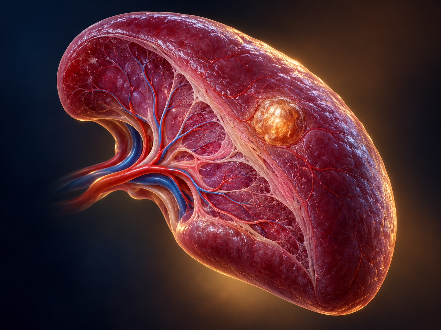
Conditions in this group
This group includes hemangioma, hamartoma, lymphangioma, inflammatory pseudotumor, primary splenic tumors, angiosarcoma, lymphoma, and splenic metastases.
How they develop and how they are diagnosed
Most focal lesions are discovered incidentally. Benign lesions, such as hemangioma, often cause no symptoms. Malignant lesions are less common and may be associated with weight loss, fever, night sweats, splenomegaly, or abnormal blood tests.
Imaging assessment with CT or MRI is central. Previous studies are compared, and a history of malignancy, the presence of lymph nodes, and blood tests are evaluated. In selected cases, PET/CT may be used or biopsy may be required. The decision to perform a biopsy is made carefully because of the risk of bleeding.
Treatment
Typical benign and asymptomatic lesions can be monitored. Surgical treatment is considered when there is diagnostic uncertainty, growth, symptoms, risk of bleeding, or suspicion of malignancy. In lymphoma or metastatic disease, treatment is usually determined in a multidisciplinary team, and splenectomy has a selected diagnostic or therapeutic role.
Frequently asked questions
Is every splenic lesion malignant?
No. Most incidental lesions are benign, but they need to be characterized correctly.
Is a biopsy always needed?
No. Imaging is often sufficient. Biopsy or splenectomy is selected only when it will safely change the diagnosis or treatment.
Can splenectomy be performed with a minimally invasive technique?
In many elective cases, yes. The choice depends on size, anatomy, and the likelihood of malignancy.
